
Abstract
In recent years, marihuana consumption has increased and become more common especially in younger patients. However, a possible association between psychosis and cannabis consumption has been described. In this paper, we present a case report of cannabis induced psychosis in a young man with a normal MRI but abnormalities in the connectivity between the caudate nucleus and frontal lobe, according to tractography imaging.
Keywords: Psychosis; Cannabis; Caudate nucleus; Frontal lobe; Tractography
Introduction
Recent studies have described a strong association between cannabis consumption and psychosis, especially in those that begin substance abuse before adulthood [1]. Cannabis, being the most common drug consumed by young adults, has an onset rate of psychosis between 35-45%. Continued consumption after an episode is associated with negative outcomes such as remission, suicidal tendencies and violence [2].
A prospective study carried out in Australia by Wade and colleagues reported a 51% chance psychotic episode in cannabis users over a 15-month period. Patients who did not consume cannabis only had a 17% chance of relapse [3]. A Dutch study reported similar results with a 42% relapse rate among persistent cannabis users compared with a 17% relapse ratein non-users [4]. Studies also described rates of psychotic episodes in a dose dependent manner. McHugh and colleagues described a 1.4-fold risk of psychotic episodes in users versus nonusers, but when compared with chronic users this risk increased up to 2.1 [5].
Despite the fact that the association between early psychotic episodes and cannabis consumption in young adults has been thoroughly studied, the precise physiopathology is yet to be studied. One theory suggests that an increase in cannabinoid CB1 receptor density in the prefrontal lobe during adolescence may be a predisposing factor [6]. One study reported that upon measuring dopamine levels during D2/D3 binding in specific radioligands, there was a 20% reduction at striatal levels in subjects exposed to cannabis [7].
Neuroimaging studies have found that prolonged use of cannabis could be responsible for a decrease in brain volume due to loss of inhibitory neurons especially in the neocortex. During the onset of psychotic symptoms there is abnormal gabanergic activity in the Anterior Cingulate Cortex (ACC), the Dorsolateral Prefrontal Cortex (DPC), and in the primary motor and visual cortex. It is thought that the absence of an inhibitory mechanism such as this could lead to the perceived cognitive dysfunction in schizophrenia [8,9].
This paper’s objective is to report a case of cannabis induced psychosis in a young man with a normal MRI but abnormalities in the connectivity between the caudate nucleus and frontal lobe according to tractography imaging.
Case Report
A 19-year-old man was referred to us and hospitalized due to visual and auditory hallucinations associated with motor restlessness after consuming cannabis. His past medical history was significant for cannabis consumption since he was 14 years.
Clinical examination revealed normal vital signs. Upon neurologic examination, a positive glabellar reflex was found along with a positive Hofmann´s sign; the rest of the examination was normal. Laboratory studies were normal. The patient was subjected to neuroimaging by MRI which was normal (Figures 1-3). Tractography revealed a loss of connectivity between the caudate nucleus and the frontal lobe as seen in (Figure 4).
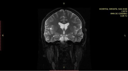
Figure 1: T2 Coronal MRI images are normal.
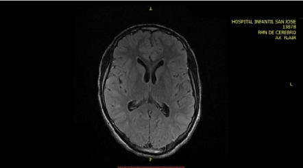
Figure 2: Flair axial MRI images are normal.
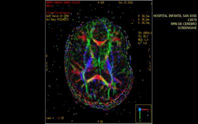
Figure 3: Anitropic map of ROI caudate nucleus.
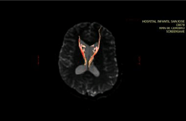
    Figure 4: Tractography shows a loss of connectivity between the caudate
nucleus and the frontal lobe.
Discussion
Studies on the endogenous cannabinoid system reveal that the psychoactive effects associated with the use of cannabis are due to the pharmacological interaction of Δ9-Tetrahydrocannabinol (THC), present in cannabis and its presentations, with one of the molecular elements in this system, the CB1 cannabinoid receptors.
The CB1 receptors, also known as central receptors, are distributed along the frontal region of the cerebral cortex, the basal ganglia, the cerebellum, the limbic system and the hypothalamus at the level of the central nervous system [10]. They are confined to the presynaptic terminals where they modulate the release of excitatory and inhibitory neurotransmitters. It is at these glutamatergic terminals that the receptors serve as an excitatory agonist with a neuro protective function and are responsible for the effects of THC on locomotion, hypothermia, analgesia and catalepsy. On the other hand, CB1 receptors that are located at GABAergic terminals are responsible for memory deficits associated with THC as well as the feeling of reward following consumption [11].
Patients diagnosed with schizophrenia, regardless of cannabis consumption, show an increase of CB1 receptor in Brodmann area 9. Cannabis consumers however, have an increase of CB1 receptors in the caudate nucleus and putamen which are part of the basal ganglia [12].
Furthermore, exposure to THC leads to an increase in the concentration of dopamine in the ventral tegmental area and in the Nucleus Acumbens (NAc) which is pivotal in the mesolimbicmesocortical pathway. This pathway is responsible for the addiction to psychoactive substances, most likely due to the inhibition of the neurotransmission of γ-aminobutyric acid (GABA). In addition, repeated long-term administration may produce the phenomenon of hypofrontality, due to the reduction of dopamine in the prefrontal cortex. Hypofrontality leads to impulsive and repetitive behavior and non-extinction of behavior. Impulsivity in particular, relates to the delay of rewards which could explain the uncontrolled urges to consume cannabis regardless of the consequences [12]. Abnormal neurotransmission of dopamine, primarily in the striatal region, is considered to be the final pathway responsible for prominent psychotic symptoms.
The acute effects of cannabinoid agonists are likely modulated by genetic factors. Several studies have explored the influence of specific genes involved in schizophrenia and their relation to the acute effects of cannabinoid psychosis. These are as follows
COMT: Catechol-O-MetylTransferase enzyme plays an important role in the decomposition of dopamine, a mutation in the COMT V gene [108/158] leads to a significant reduction in the activity of this enzyme. Patients who have COMT polymorphisms have a moderate risk of developing psychotic symptoms.
DAT1: in the striatum, synaptic dopamine clearance is dependent on this dopamine transporter, a polymorphism of the DAT1 gene is associated with low transport and high levels of dopamine in the striatum. Bhattacharyya and colleagues described that in individuals with one allele and 9 replicates had higher risk for THC-induced psychotic symptoms.
AKT1: inactivates glycogen synthase kinase by phosphorylation and has been associated with schizophrenia. Cannabinoids stimulate the AKT1 pathway through CB1 receptors. Bhattacharyya and colleagues also found that a polymorphism in the AKT1 gene [the GG genotype of a single nucleotide polymorphism rs1130233] induces moderate sensitivity to acute psychotic episodes by THC.
BNF: studies have found that patients who have polymorphisms in these genes and begin cannabis consumption at an early age are more susceptible to having a psychotic episode [13].
Conclusion
The clinical presentation of this patient may be explained by the compromise of connectivity between the caudate nucleus head and the frontal lobe, considering that this connectivity is dopamine mediated and that different cannabinoid receptors may modulate dopamine’s receptors.
References
- Mane A, Bergé D, Penzol MJ, Parellada M, Bioque M, Lobo A, et al. Cannabis use, COMT, BDF and age at first-episode psychosis. In Psychiatry Research. 2017; 250: 38-43.
- Lambert M, Conus P, Lubman DI, Wade D, Yuen H, Moritz S, et al. The impact of substance use disorders on clinical outcome in 643 patients with first-episode psychosis. Acta Psychiatr Scand. 2005; 112: 141-148.
- Wade D, Harrigan S, Edwards J, Burgess PM, Whelan G, McGorry PD. Substance misuse in first-episode psychosis: 15-month prospective followup study. Br J Psychiatry. 2006; 189: 229-234.
- Linszen D, Dingemans P, Lenior M. Cannabis abuse and the course of recentonset schizophrenic disorders. Arch Gen Psychiatry. 1994; 51: 273-274.
- McHugh MJ, McGorry PD, Wood SJ, Hartmann JA, Nelson B, Yung AR, et al. Cannabis-induced attenuated psychotic symptoms: implications for prognosis in young people at ultra high risk for psychosis. Psychological Medicine, 2017; 47: 616-626.
- Caballero A, Tseng KY. Association of cannabis use during adolescence, prefrontal CB1 receptor signaling, and schizophrenia. Front Pharmacol. 2012; 3: 101-103.
- Sami MB, Rabiner EA, Bhattacharyya S. Does cannabis affect dopaminergic signaling in the human brain? Asystematic review of Evidence to date. Eur Neuropsychopharmacol. 2015; 25: 1201-1224.
- Hashimoto T, Bazmi HH, Mirnics K, Wu Q, Sampson AR, Lewis DA. Conserved regional patterns of GABA-related transcript expression in the neocortex of subjects with schizophrenia. Am J Psychiatry. 2008; 165: 479- 489.
- Rais M, Cahn W, Van Haren N, Schnack H, Caspers E, Hulshoff PH, et al. Excessive brain volume loss over time in cannabis-using first-episode schizophrenia patients. Am J Psychiatry. 2008; 165: 490-496.
- Escobar TE, BerrouetMC, Gonzalez DM. Mecanismos moleculares de la adicción a la marihuana. Rev Colomb Psiquiat. 2009; 38.
- Maldonado R, Berrendero F. Neurochemical Basis of cannabis addiction. Neuroscience. 2011; 181; 1-17.
- Sofía Tziraki. Trastornos mentales y afectación neuropsicológica relacionados con el uso crónico de cannabis. RevNeurol. 2012; 54: 750-760.
- SherifM, Radhakrishnan R, D’Souza D, Ranganathan M. Human Laboratory Studies on Cannabinoids and Psychosis. Biological Psychiatry. 2016; 79: 526-538.