Case Report
Austin J Anat. 2015; 2(3): 1039.
Unusual Branching Pattern of Left Coronary Artery- Case Report
Rajani S*
Department of Anatomy, All India Institute of Medical Sciences, India
*Corresponding author: Rajani Singh, Department of anatomy, All India Institute of Medical Sciences, India
Received: September 05, 2015; Accepted: September 15, 2015; Published: November 02, 2015
Abstract
During dissection of thorax of an 80 year old Female cadaver for undergraduate MBBS students, a heart was detected to have variant branching pattern of left coronary artery. Typically left coronary artery bifurcates into anterior interventricular artery and circumflex artery which continues in the left coronary sulcus. But the present case left coronary artery splitted into anterior interventricular artery and common trunk. The anterior interventricular artery gave diagonal/ventricular branches in the left as usual but ectopically generated a ventricular minor artery and a luminal artery. The luminal artery entered the infundibulum of right ventricle and ended near the septal papillary muscle. Common trunk divided into circumflex artery and anomalous diagonal artery. The case is reported for its virgin occurrence.
The knowledge of new configuration of branches of left coronary artery will be of paramount importance to cardiologists in cardiac surgery and to anatomists for new variant.
Keywords: Circumflex artery; Infundibulum of right ventricle; Left coronary artery; Luminal artery; Ventricle minimus artery
Abbreviations
LCA: Left Coronary Artery; RCA: Right Coronary Artery; AIV: Anterior Interventricular Artery; Cx: Circumflex
Introduction
Heart is supplied by two coronary arteries, Left Coronary Artery (LCA) and Right Coronary Artery (RCA). Variations in the branching pattern of LCA have been described by many authors [1, 2].
Typically LCA gives Anterior Interventricular Artery (AIV) and main artery continues as circumflex artery in left anterior coronary sulcus. Anterior interventricular artery from its left side gives many sub-parallel diagonal and ventricular branches to supply the left ventricle. But in the present cadaveric study, two arteries named as ventricular minor artery and luminal artery ectopically arise from right side of AIV. Ventricular minor artery enters the myocardium and ends there. Luminal artery enters into the lumen of right ventricle of heart.
In addition to these new arteries, a diagonal artery arises from common trunk along with circumflex artery. These variations are not reported in literature as far as known to the author and may affect the pathophysiology of heart. So the knowledge of these variants may be of immense use to clinicians, imagery experts and anatomists. Therefore the case is reported.
Case Report
During dissection of thorax of an eighty year old female cadaver, the heart was found to have variant configuration of branching pattern of LCA as well as AIV as described below-
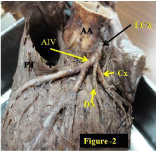
LCA after travelling 1.5 cm from its origin gave a common trunk of 1.00 cm and AIV (Figure 2). Common trunk bifurcated into Circumflex (Cx) and diagonal arteries. Cx artery after travelling 2.00 cm in the left anterior coronary sulcus gave marginal artery and ended aberrantly at the left border of heart.

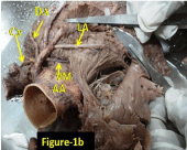
    Figure 1b:  shows extent of luminal artery in the cavity of right ventricle after
opening the right ventricle.
VM: Ventricular Minor; LA: Luminal Artery; Cx: Circumflex Artery; DA:
Diagonal Artery; AA: Abdominal Aorta.

    Figure 2:  Common trunk giving circumflex and diagonal arteries.
LCA: Left Coronary Artery; Cx: Circumflex Artery; DA: Diagonal Artery; AIV:
Anterior Ventricular Artery.
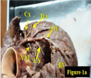
A new artery named as ventricular minor (Figure 1a and 1b) arose from right side of AIV at 0.50 cm from its origin. The diameter of this artery was 0.15 cm and length was 3.00 cm. This artery, after its emergence, entered into the myocardium of right ventricle and ended there. Further on the same side, another new artery named as luminal artery arose from the AIV after 1.50 cm from its origin. This artery passed between the ascending aorta and pulmonary trunk and then entered the infundibulum of right ventricle (Figure 1a). Travelling through the infundibulum, it ended at the septal papillary muscle of the right ventricle (as exposed after opening the ventricle Figure 1b). The luminal artery after travelling for 2.00 cm having uniform diameter of 0.25 cm in the right ventricle gave a small arterial branch. Running further for 1.10 cm gave another branch and ended in the papillary muscle with reduced diameter of 0.08 cm. All the measurements were taken by venire caliper.

    Figure 1a:  Luminal artery entering the infundibulum.
VM: Ventricular Minimus; LA: Luminal Artery; Cx: Circumflex Artery; DA:
Diagonal Artery; AIV: Anterior Ventricular Artery; RV: Right Ventricle.
Discussion
According to some authors, normal variant is an alternative pattern which is relatively infrequent compared to normal, but it occurs in more than 1% of otherwise normal individuals [3, 4]. The advances made in coronary arterial bypass surgeries and modern methods of myocardial revascularization makes sound and complete knowledge of the normal and variant anatomy of coronary artery [5] indispensable and imperative. Diagonal branch normally arises from AIV/Cx arteries as described in Gray’s anatomy (2008) [6] but in this case common trunk gives diagonal artery and Cx artery. Ectopic luminal artery too arises from AIV and entering in the lumen of right ventricle. That is why it has been named as luminal artery. This luminal artery is a new discovery and being reported for the first time. The diameter of this artery is uniform showing that it is not affected by luminal blood and there was no differential influence of externally surrounding blood flow on the uniform width of the artery. Widening of blood vessel occurs due to sheer flow of blood through them [7]. Uniform diameter of luminal artery suggests that it must have accommodated the blood during fetal life time. The cause of the formation of luminal artery may be developmental, genetic or circumstantial. It is speculated that the normal supply to septal papillary muscle might have been insufficient during developmental stage. Therefore luminal artery might have been generated. The discovery of luminal artery may change traditional old concepts of supply to papillary muscle.
References
- Baptista CA, Didio LJ, Prates JC. Types of division of the left coronary artery and the ramus diagonalis of the human heart. Japanese heart journal. 1991; 32: 323-35.
- Lujinovic A, Ovcina F, Voljevica A, Hasanovic A. Branching of main trunk of left coronary artery and importance of its diagonal branch in cases of coronary insufficiency. Bosn J Med. 2005; 5: 69-73.
- Angelini P. Normal and anomalous arteries- Definitions and classification. Am Heart J. 1989; 117: 418-434.
- Angelini P. Coronary artery anomalies-Current clinical issues: Definitions, classifications, incidence, clinical relevance and treatment guidelines. Tex Heart Inst J. 2002; 29: 271-278.
- Dombe DD, Anitha T, Giri PA, Swapnali D. Dombe, Medha V, et al. Clinically relevant morphometric analysis of left coronary artery. Int J Biol Med Res. 2012; 3: 1327-1330.
- Standring S. Gray’Anatomy: The anatomical basis of clinical anatomy 40th Edition. London Churchill Livingstone: Elsevier. 2008; 980.
- Jones EAV, Le Noble F, Eichmann A. What determines blood vessel structure? Genetic prespecification vs. hemodynamics. Physiology. 2006; 21: 388-395.