Case Report
Austin J Anat. 2015; 2(3): 1040.
Unusual Position and Size of the Appendix and the Sigma Colon
Maslarski I*
Department of Anatomy, histology and pathology, University of Medical Faculty at the Sofia, Bulgaria
*Corresponding author: Maslarski I, Department of Anatomy, histology and pathology, University of Medical Faculty at the Sofia, Bulgaria
Received: August 10, 2015; Accepted: August 20, 2015; Published: November 13, 2015
Abstract
The topography of the large intestine and the additional elements, like the appendix, are well known, but their variants deserve further attention. During our academic dissection of the cadaver, completed with medical students, we encountered unusual length of the Vermiform Appendix (VA), as well as abnormal size, position and curves of the descending and Sigmoid Colon. The position of the VA was retroperitoneal and retrocaecal. The size was 16.3 cm. The topography of colon parts also showed peculiarities, which differed from the norm. After the left colic flexure, the descendent colon was directed down and close to the left portions of the small intestine. Instead of moving curve less to the lower left abdomen, the colon was directed obliquely and to the right, and passed to the caecum. The observed position of the large intestine led us to the description of its topographic classification, embryonic origin and attaching apparatus. This unusual move of the descendent colon and the subsequent sigma makes the case interesting for the abdominal surgical practice.
Keywords: Vermiform appendix; Sigmoid colon; Mesentery
Introduction
The topography of the large intestine and the additional elements, like the appendix, are well known, but their variants deserve further attention. The most common variants are observed in the length and positions of the appendix. The topography of the vermiform appendix depends on the neighbor organs and the attaching apparatus of the peritoneum (mesoappendix). The main variants are Retrocaecal, Subcaecal, Paracaecal, Post Ileal, Pelvic and Preileal positions. If a patient was to have some of the listed variants, he/she normally would not feel any specific symptoms. But inflammation of an appendix positioned abnormally may imitate diseases, like: colits, pelvic inflammatory disease, sub hepatic – hepatitis, torsion of the ovarian cyst, etc. The Vermiform Appendix (VA) has the role of a tonsil. Both of these structures are composed of lymphatic tissue. The appendix in surgical practice is detected by tracing the three longitudinal muscle layers, whose terminal fusion composes the proximal part of the gut extension. The growth of longitudinal muscle layers (the taeniacolli) may confuse the surgeon about the real position of the appendix.
The Sigmoid Colon is the terminal section of the large intestine and is situated in the pelvic region. The anterior part of the colon is separated from the bladder in the male, and from the uterus in the female. The external iliac vessels, the left sacral plexus and the left piriformis muscle are situated posterior the sigmoid colon. Diverticulitis often occurs in the sigmoid colon. The unusual size, topography and relationships were documented.
Case Presentation
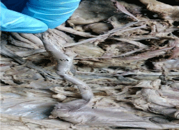
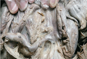
During our academic dissection of the cadaver, completed with the help of medical students, we encountered an unusual length of the Vermiform Appendix (VA), as well as an abnormal size, position and curves of the descendent and Sigmoid Colon. The medical students were from the Department of Anatomy, Histology and Pathology, at the Medical Faculty of the University of Sofia. The cadaver belonged to a 70-year-old woman and was fixed with the formaldehyde method. When observing the abdominal organs and their positions, we discovered the unusual position and size of the VA. The position of the VA was retroperitoneal and retrocaecal. The curve of the VA was extreme, and passed through the ascending part of the colon and against the liver. The size of the VA was 16.3cm (Figure 1). Typically the caecum is located intra peritoneally in the lower right abdomen and has a length of 5 to 7 cm. The VA attached itself with a double peritoneal layer to the back of the abdominal wall, becoming the mesoappendix (Figure 2). Here taeniae, haustra and semi lunar folds were all absent. The diameter of the VA was approximately 0.8 cm.

    Figure 1:  Unusual length of the vermiform appendix.
VA: Vermiform Appendix; C: Caecum

    Figure 2:  Interactions between parts of the mesoappendix.
VA: Vermiform Appendix; C: Caecum; MA: Mesoappendix Attachment
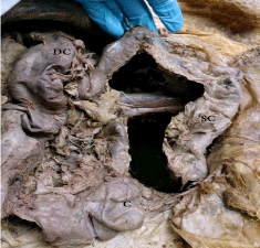
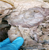
The topography of the parts of the colon also showed peculiarities, which differed from the norm. After the left colic flexure, the descending colon was directed downwards and near to the left portions of the small intestine. Instead of moving curveless to the lower left abdomen, the colon was directed obliquely and to the right, and passed to the caecum. This unusual position of the descending colon and the subsequent sigma makes the case interesting for the abdominal surgical practice (Figure 3). The Sigmoid Colon carried its typical curvature to the caecum, while the serosa of the above mentioned parts of the colon were in direct contact. Their separation severed a portion of the anchoring device (Figure 4). After this contact, the Sigmoid Colon deviated to the left and entered the pelvis, as opposed to the typical procedure of entering from the lower left abdomen. In the transition area between the small and the large intestines, there was also an interesting attachment apparatus between the caecum, the appendix and the Sigmoid Colon.

    Figure 3:  Topography of the descending colon and the sigma colon.
DC: Descending Colon; SC: Sigma Colon; C: Caecum

    Figure 4:  Contact between the sigma colon and the caecum.
SC: Sigma Colon; C: Caecum; SA: Serosa Attachment; SI: Small Intestine
Discussion
In order to observe the position of the large intestine, one must be aware of the embryonic origin, the attaching apparatus and the topographic classification of the organ. In this case report, we observed the length and the position of the Vermiform Appendix (VA) and the Sigmoid Colon.
According to Wakeley’s [1,2] classification of the VA, the variations in its position and pathway could be: retrocaecal, retroperitoneal (passing behind the cecum and the ascending colon), pelvic, splenic or ileal, subcecal and paracolic, and mid inguinal. In this case report, we observed the retrocaecal and the retroperitoneal type VA, the latter possessing an abnormally big mesoappendix. O’Connell’s classification provides a more detailed picture of the retrocaecal VA subgroups using its topographic position. According to him, our case would be the 3rd type of VA adherent to the posterior surface of the caecum and the ascending colon. The size of the VA in our case was more than 16 cm and the position was closer to the liver.
We have referred to anatomy atlases and topographic literature in order to determine the typical topographical position of the Sigmoid Colon. According to Hafferl A. [3,4], the author of “Topographical Anatomy”, the Sigmoid Colon is in an abnormal position, when bulges on the outside of the intestine, called diverticula, are formed due to the inner, soft tissue, layer of the colon pushing through the outer, muscular layer. The colon loop of the Sigmoid Colon then reaches the right iliac fossa. Furthermore, Netter’s Anatomy Atlas provided us with a name for this intestine position: “loops to the right side of the colon” sigmoid [5]. Position variations of the colon and the mesoappendix can be explained by the embryonic development. The gut tube is the embryo structure; as such, it is the precursor of the respiratory and digestive systems and is derived from the endoderm and the mesoderm in human development [6]. This structure is subdivided into the foregut, midgut and hindgut. The midgut generated the intestine subdivisions and portions of the large intestine, including the caecum and the VA. The hindgut consisted of the large intestine, including the Sigmoid Colon. The splanchnic mesoderm, adjacent to the endoderm, formed a sheet of tissue, which enclosed the gut tube (primitive gut). This sheet of tissue was the common Mesentery and was attached to the dorsal and ventral body walls along the medial line. The space between the intestine and the dorsal body wall was the dorsal Mesentery and was relevant to our case. Under “Mesentery” we understand “a fold of tissue that attaches organs to the body wall”. It usually refers to the small intestine, but it also exists to support the large intestine, such as the Sigmoid Colon or the appendix. As mentioned earlier in this case report, the mesoappendix of the appendix and the sigma colon were in close contact. The contact can be explained with the common embryonic origin of these Mesentery structures.
Variations in the length and position of the VA and of the Sigmoid Colon may lead to some pathological conditions. The redundant loop of the Sigmoid Colon can cause various problems such as indigestion, insomnia, pain in the right iliac fossa or loss of weight. Some cases [7] are known for the excessively long distal part of the colon, including the right-sided Sigmoid Colon. There is some variation in the position of the Sigmoid Colon in adults, however, as the radiographs show [8]. (It was noticed that, on radiographs of young children, the Sigmoid Colon was often positioned within the right lower quadrant, as opposed to the left lower quadrants in adults). The Sigmoid Colon in this case could be mistaken for the caecum or the ascending colon and lead to false diagnosis.
The description of this case could improve diagnostic activity during the radiological studies and be beneficial for future surgical practice.
References
- Geethanjali H, Lakshmi P. A Study of Variations in the Position of Vermiform Appendix, Anatomica Karnataka. 2011; 5: 17-23.
- Wakeley CP. The position of the vermiform appendix as ascertained by analysis of 10000 cases. J Anat. 1933; 67: 277-283.
- Hardin DM. Acute Appendicitis: Review and Update, Am Fam Physician. 1999; 60: 2027-2034.
- Hafferl A. Lehrbuch der topographischen anatomie, Springer – Verlag, Berlin.
- Floch M. Netter's Gastroenterology, 2nd edition. Saunders Elsevier, Philadelphia. 2009; 367-370.
- Moore K, Persaud T. Before we are born- Essentials of embryology and birth defects. Saunders Company, Philadelphia. 1998; 273-280.
- Nayak SB. George BM, Mishr S. Abnormal Length and Position of the Sigmoid Colon and Its Clinical Significance. 40; 2012; 10: 94-97.
- Prassopoulos P, Gourtsoyiannis N, Cavouras D, Pantelidis N. Interposition of the colon between the kidney and the psoas muscle: a normal anatomic variation studied by CT, Abdominal imaging, Springer – Verlag, New York. 1994; 19: 446- 448.