Abstract
Coronary Artery Disease (CAD) is a leading cause of death worldwide. Early detection has been shown to be critical in preventing CAD-related deaths. Magnetocardiography (MCG) is often favoured for its non-invasiveness and high sensitivity in the current diagnosis of CAD. Despite the popularity of MCG, an analysis of its cost-effectiveness in comparison with other non-invasive methods has not yet been performed. To estimate the potential cost effectiveness of MCG in CAD patients, specifically in those with chest pain, cost-effectiveness analyses of selected non-invasive methods (Stress-ECG, Stress-Scintigraphy and Stress-EchoCG) were performed and compared. The analysis revealed that MCG shows the lowest cost-effectiveness ratios, indicating it is the most efficient diagnostic method amongst non-invasive cardiographs. Furthermore, our analysis revealed that MCG is the most cost efficient method even for patients with symptomatic indication of CAD (e.g. chest pain), either on its own or in combination with coronary angiographs. These results suggest MCG is a highly most economical non-invasive diagnostic method, and can improve the quality of CAD diagnosis.
Keywords: Magnetocardiography; Cost-effectiveness analysis; Coronary artery disease; Non-invasive diagnostic methods
Abbreviations
MCG: Magnetocardiography; CAD: Coronary Artery Disease; CEA: Cost-Effectiveness Analysis; Stress-ECG: Stress Electrocardiography; Stress-scintigraphy: Stress Scintigraphy Test with Tallium; Stress-EchoCG: 2D Echocardiography and Load Test with Treadmill; RISK: Risk of Essential Cardiovascular Failures Provoked by Given Diagnostic Method; CA: Coronary Angiography; CER: Cost-Effectiveness Ratio; PREV: Prevalence; SENS: Sensitivity; SPEC: Specificity
Introduction
Cardiovascular complications represent one of the leading cause of death worldwide, and they are estimated to cause 23.3 million deaths by 2030 [1]. Coronary Artery Disease (CAD) is the most common cause of death among cardiovascular complications, and indeed it accounted for more than 16.8% of all deaths worldwide in 2013 [2]. CAD has also been associated with important morbidity and mortality related to stroke, ischemia, embolism and heart failure [3]. The number of cases of CAD is especially high in developed countries. According to the Global Burden of Disease Study 2010, ischemic heart disease and stroke are the most prevalent diseases in Ukraine. In the United States, CAD is the most common cause of death in men and women over 20 years of age, contributing to 370,000 deaths annually [4].
Especially concerning is the fact that the prevalence of CAD is increasing [5]. Moreover, the identification of the mechanisms by which CAD results in untimely deaths, as well as the development of safe and effective therapies to combat it, remain elusive. Developing innovative therapeutics targeting CAD is a priority, and much effort has been expended in identifying prophylactic measures and pharmacological approaches for disease management. While the methods of early CAD diagnosis have been significantly improved, stress echocardiography, followed by Coronary Angiograph (CA), remain the most favourable methods of diagnosis in symptomatic patients. However, the recent development of Magnetocardiography (MCG) - a non-invasive cardiac-activity mapping technique - has led to increased detection sensitivity via increased numbers of recording sites as compared to other non-invasive cardiographs. MCG can detect even slight changes in the electrophysiology of the myocardium, and allows for the visualization of cardiac electrophysiological processes without any external influence [6]. MCG also provides information on the magnetic signature produced by the vortex currents in the myocardium, which cannot be registered by Electrocardiography (ECG) [7]. These unique advantages of MCG make it an attractive technique for CAD detection and it has contributed to the current understanding of the generation, localization, and dynamic behaviours of cardiac currents in CAD patients.
Common non-invasive techniques to diagnose CAD include Stress Induced Electrocardiography (stress-ECG), Echocardiography (stress-EchoCG) and Scintigraphy (stress-scintigraphy). The choice of one method over another depends on cost-effectiveness and resource consideration. Generating a generic model that can estimate the comparative cost effectiveness of a screening technique would thus provide a valuable tool to assess the opportunity cost of a medical intervention on the health care system [8]. In order to make a comparison amongst the current non-invasive cardiographs, this study aimed to perform a Cost Effectiveness Analysis (CEA) for several methods (Stress-ECG, Stress-EchoCG, and Stress-Scintigraphy) and to compare them with the MCG, the most modern non-invasive CAD diagnostic technique.
Methods
Statistical definitions
The research was conducted under the following terms:
- Prevalence (PREV) of CAD: estimated to be 10% in this theoretical model [9].
- α = 1 – Sensitivity (SENS): represents the probability of the diagnostic method correctly detecting patients with CAD.
- β = 1 – Specificity (SPEC): represents the probability of the diagnostic method correctly detecting patients without CAD.
Total probability of false diagnosis for CAD
Health economic evaluations include uncertainty for both positive and false parameters of observable variables. In order to determine the total probability of establishing a false diagnosis, we have built a generalized model to combine both sensitivity and specificity to analyze the likelihood of an error [10]:
Total probability of false diagnosis for CAD in patients with symptomatic indications
The most common symptom of CAD is chest pain, described as chest discomfort, aching, and heaviness in the chest [11]. Since the presence of disease symptoms can bias the selection of a method of diagnosis, prevalence – which measures the probability of a randomized occurrence of CAD - is not an accurate measurement in our model. Pretest Probability (PP), determined by the Mayo Clinic Index (MCI) from 2002, was used instead to calculate the total probability of false diagnosis of CAD in patients with chest pain [12].
Cost-effectiveness ratio
Diagnostic accuracy incorporates parameters of Specificity (SPEC), Sensitivity (SENS) and Prevalence (PRV) when measuring the effectiveness of a method. To determine the diagnostic accuracy of non-invasive methods used in medical practice for the diagnosis of CAD, we calculated predictive indexes using values of sensitivity and specificity derived from the literature [13,14]:
where NPV is a negative predictive value (rate of coincidence of negative test results under the absence of CAD); PPV is a positive predictive value (rate of coincidence of positive test results under the presence of CAD); SPEC and SENS - specificity and sensitivity, respectively; PREV - the prevalence of CAD; α and β -probability of the diagnostic method correctly detecting patients with or without CAD, respectively.
Based on formulas 3 and 4, the average of the diagnostic effectiveness was calculated [13,14]:
The Cost-Effectiveness Ratio (CER) of each diagnostic method was also calculated:
Substituting the calculated value of effect (5) above, we can reformulate the CER as the following:
Ratio for cost-effectiveness increments
The Incremental Cost-Effective Ratio (ICER) provides the summarized cost-effectiveness of a health care intervention by comparing the CER of two diagnostic methods. For this analysis, we have calculated the coefficient ratio between the MCG and other noninvasive cardiographs using the following equation:
where ICER is the incremental cost-effectiveness ratio; Cost(MCG) is the relative cost of MCG; Cost(NIM) is the relative cost of another non-invasive method; Effect(MCG) is the diagnostic accuracy of MCG; Effect(NIM) is the diagnostic accuracy of another non-invasive method.
Relative cost of non-invasive diagnosis followed by coronary angiography
The accuracy of a non-invasive method to diagnose CAD can be uncertain due to the sensitivity and specificity of the method, as well as the severity of the disease symptoms. In most cases, 70% to 90% of diagnoses using non-invasive methods still require a CA to fully confirm the presence of CAD. Therefore, we have further modified the generated formula to calculate the relative probable cost for patients with or without CAD when undergoing both invasive and non-invasive diagnostic methods.
The relative cost of a false diagnosis for patients without symptomatic implications was calculated using:
The relative cost of a false diagnosis for patients with symptomatic implications was calculated using:
where PREV is the prevalence of CAD; PP is the pretest probability of CAD based on symptomatic indication; α and β are the probability of the diagnostic method correctly identifying patients with or without CAD, respectively; α(CA) and β(CA) (both = 0.001) are the probability of false positive and negative diagnosis, respectively [15]; Cost (CA) is the cost of the coronary angiograph.
Results
Total probability of false diagnosis for CAD using selected non-invasive methods
To establish the overall effectiveness of selected medical interventions, both diagnostic accuracy and cost were evaluated for the purpose of this study. The cost-effectiveness of MCG was compared with that of other non-invasive methods, namely Stress- ECG, Stress-scintigraphy and Stress-EchoCG, to determine which method is the most accurate with the lowest cost. Sensitivity (SENS) and Specificity (SPEC), derived from previously reported analyses [13,14], and the relative costs of examinations were compared in Table 1.
    Diagnostic method
    Sensitivity (SENS)
    Specificity (SPEC)
    Risk
    Relative cost of examination (Cost)
    Reference


    MCG
    0.93
    0.84
    0.0 %
    1
    [15]


    Stress-ECG
    0.68
    0.77
    0.05%
    0.8
    [16]


    Stress-SCN
    0.90
    0,77
    0.05%
    3.3
    [16]


    Stress-EchoCG
    0.84
    0,87
    0.05%
    2.5
    [16]


    CA
    0.99
    0.99
    1.5 %
    7.8
    [16]
Table 1: Statistical Measures, Risks and Relative Costs for CAD diagnostic methods.
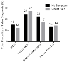
To include the possibility of a false diagnosis, the total probability of a false diagnosis for each non-invasive method was first calculated from the generated model above (1, 2) (Figure 1).

    Figure 1:  Total probability of false diagnoses for CAD by non-invasive
methods.
We observed that the stress induced ECG method (Perror = 24%) resulted in the highest probability of false diagnosis, while stress induced EchoCG exhibited the lowest (Perror = 13%). When symptoms of chest pain were present, MCG showed the lowest probability of false diagnosis in comparison to other non-invasive diagnostic methods (Perror = 15%). The probability of false diagnosis between stress induced EchoCG and MCG was not statistically different, indicating that the two methods have a similar rate of misdiagnosis.
Relative costs of false diagnosis using selected noninvasive methods in combination with coronary angiography
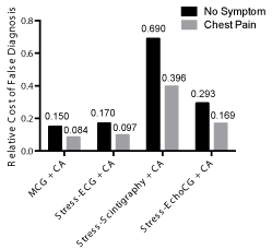
Conventional X-ray CA is the standard of reference for the assessment of CAD. The ability of a CA to detect both the exact location of CAD as well as the severity of the disease makes it an attractive method to confirm CAD diagnosis. Therefore, even after the usage of a non-invasive method, a CA is often performed to validate the diagnosis. Although we observed that MCG has the lowest probability of false diagnosis, the relative cost of an individual diagnosis using MCG is neither accurate nor realistic in current medical practice, as CA is often used in conjunction with a non-invasive technique. Therefore, we conducted a further analysis comparing the relative cost of false diagnosis using non-invasive diagnostic methods in combination with CA (Figure 2).

    Figure 2:  Relative costs of false diagnosis of CAD using a combined noninvasive
method and CA.
ECG: Electrocardiography; EchoCG: Echocardiography; CAD: Coronary
Artery Disease; MCG: Magnetocardiography; CA: Coronary Angiography.
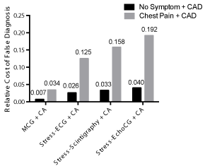
Our assessment revealed that the MCG + CA diagnostic combination has the lowest relative cost of false diagnosis (0.145). The stress induced ECG + CA combination exhibited the second lowest cost, (0.167), making it 15% more expensive than MCG + CA. Stress-scintigraphy + CA had the highest relative cost of false diagnosis (0.685), making the cost 370% greater than MCG + CA. In patients with CAD, the relative costs of false diagnosis were found to be minimal, yet MCG still exhibited the lowest relative cost amongst the non-invasive methods (Figure 3).

    Figure 3:  Relative costs of false diagnosis in patients with chest pain using
combined non-invasive method and CA.
ECG: Electrocardiography; EchoCG: Echocardiography; CAD: Coronary
Artery Disease; MCG: Magnetocardiography; CA: Coronary Angriography.
When patients presented with symptomatic indications of CAD (e.g. chest pain), patients without CAD had relatively lower costs of false diagnosis than patients without symptoms. Furthermore, symptomatic indications did not alter the overall trend; only the relative cost of false diagnosis was decreased. On the other hand, patients with CAD had increased relative costs of false diagnosis when presented with chest pain. This is expected, as CA is often the commonly used diagnostic method, and thus having additional non-invasive procedures is considered unnecessary and only adds additional costs.
Cost-effectiveness analysis (CEA) for non-invasive diagnostic methods of CAD
Incremental cost-effectiveness ratios amongst the non-invasive diagnostic methods of CAD were used to make direct comparisons of each method’s cost effectiveness (Table 2). From our generated model, MCG and stress-ECG had cost-effectiveness ratios of 1.4 and 1.3, respectively. Stress-EchoCG had a cost effectiveness ratio of 3.6, and stress-scintigraphy exhibited the highest ratio of 5.1. We next compared the ICER value of MCG to those of other non-invasive CAD diagnostic methods, and found all the ratios generated by this comparison were greater than 1 (ICER > 1). An ICER value greater than 1 indicates that the difference in the diagnostic effectiveness of MCG versus other non-invasive methods is lower than the difference in their costs, thus demonstrating that MCG is the most cost-effective diagnostic method amongst those studied.
    Diagnostic method
    Predictive value
    Effectiveness
    Relative Cost
    Cost-Effectiveness Ratio, CER
    ∆ Cost= Cost(MCG)– Cost
    ∆ Effect =Effect(MCG) – Effect2
    ICER


    PPV
    NPV


    MCG
    0.39
    0.99
    0.69
    1
    1.4
    -
    -
    -


    Stress-ECG
    0.25
    0.96
    0.605
    0.8
    1.3
    0.20
    0.09
    2.40


    Stress-SCNT
    0.30
    0.99
    0.645
    3.3
    5.1
    -2.30
    0.05
    -51.1


    Stress-EchoCG
    0.42
    0.98
    0.70
    2.5
    3.6
    -1.50
    -0.01
    150
Table 2: Comparison of cost-effectiveness ratios among non-invasive diagnostic methods of CAD.
Discussion
Clinical economic analyses are necessary to justify healthcare costs. Indeed, medical decisions must now factor in healthcare costs in addition to clinical considerations. Marginal costs - the costs of providing an additional unit of service - for each medical diagnosis need to be carefully considered before an assessment is initiated. In the case of CAD, advances in the technology of non-invasive coronary artery imaging devices have improved early detection of subclinical cases. However, a comprehensive model that compares the cost-effectiveness of each non-invasive method has not been previously reported. The analysis presented in this study focused on comparing the cost-effectiveness and the relative cost of false diagnosis for each non-invasive method. Simple diagnostic analyses of the economic consequences of health benefits over cost, however, require a number of assumptions, and for this reason these analyses are rarely straight forward. In order to create a comprehensive model that compares the effectiveness of each medical diagnostic method, we have formulated a number of generic models that incorporate the following essential aspects: medical - characterized by the accuracy of the treatment, diagnosis, and frequency of rehabilitation; economical - measured by the financial medical cost and the opportunity cost for rehabilitation; and sociality - assessed by the patient’s quality of life after the treatment.
Our analysis demonstrates that under most assumptions, MCG is the most cost-effective non-invasive diagnostic method for CAD. While MCG has a higher cost than stress-ECG, it is more accurate and effective, thus overall making it a better, more cost-effective diagnostic tool. The probability of a false diagnosis using MCG was the lowest among other non-invasive procedures. Lastly, a quantitative overview of the Incremental Cost-Effectiveness Ratio (ICER) indicated that other non-invasive methods have a ratio greater than 1 in comparison to MCG. These evaluations suggest that MCG has the most optimal and practical benefits in relation to its cost. Other non-invasive methods, such as stress induced EchoCG (ICER = 150), however, are not recommended due to their low practicality and high costs.
Further analysis using a combination both non-invasive and invasive methods, specifically CA, were performed to compare relative costs of false diagnosis. CA is the most standard test for identifying the presence and extent of atherosclerotic CAD, and therefore, it is often implemented in combination with a non-invasive method. Our results indicate that in the case of false diagnosis, the highest relative cost for combination therapy occurs with stressinduced scintigraphy and CA. MCG and CA combination diagnostic methods, on the other hand, exhibited the lowest relative cost of false diagnosis (> 4 fold less than stress-scintigraphy + CA). The overall relative costs for false diagnosis were lower when patients had CAD symptomatic indications than when they did not. However, the trend was the same whether the patients had symptoms of CAD or not; stress-scintigraphy had the highest cost, while MCG had the lowest. These results illustrate that the MCG is the least expensive method when used in conjunction with CA, suggesting it should be the first line of diagnosis for CAD.
Cost-effectiveness analyses have strengths and limitations. The limitations of our study include the generalized assumption that the effectiveness of a diagnostic method can be quantified by the number of successfully identified clinical cases it detects. We did not consider any restrictions and weaknesses of each diagnostic method, including motion artifacts and soft tissue attenuation. Possible side effects, complications, and risks, involving, for example, the exposure to radiation with CA, were not included in the analysis. Therefore, a more sophisticated approach would have been generated if references to these costs were available. Nevertheless, CEA provides an overall comparison of the net benefit of each medical diagnosis. This method of analysis has become the most commonly used metric of health impact and is often applied by the World Health Organization (WHO) for their evaluations [16].
Our findings may provide crucial clinical considerations for health care providers, as they are frequently presented with an array of diagnostic methods. Cost-benefit analysis is therefore highly useful in implementing medical diagnostic techniques that are most costeffective for both patients and public health officials. Our results demonstrate that the MCG has considerably lower cost-benefit ratios in comparison to other non-invasive methods, and the high accuracy and non-invasive properties of MCG makes it the most attractive noninvasive method to diagnose CAD under current clinical parameters.
References
- Who.int,'WHO | Cardiovascular Diseases (Cvds)'. N.p. Web. 2015.
- GBD 2013 Mortality and Causes of Death Collaborators. Global, regional, and national age-sex specific all-cause and cause-specific mortality for 240 causes of death, 1990-2013: a systematic analysis for the Global Burden of Disease Study 2013. Lancet. 2015; 385: 117-171.
- Cuadrado-Godia E, Ois A, Roquer J. Heart Failure in Acute Ischemic Stroke. Current Cardiology Reviews. 2010; 6: 202-213.
- Xu J, Murphy SL, Kochanek KD, Bastian BA, Statistics V. National Vital Statistics Reports. Deaths: Final Data for 2013. 2016; 64.
- Moran AE, Forouzanfar MH, Roth GA, Mensah GA, Ezzati M, Flaxman A, et al. The Global Burden of Ischemic Heart Disease in 1990 and 2010: The Global Burden of Disease 2010 Study. Circulation. 2014; 129: 1493-1501.
- Tolstrup k. Comparison of Resting Magnetocardiography with Stress Single Photon Emission Computed Tomography in Patients with Stable and Unstable Angina. American College of Cardiology.
- Chaikovsky I, Boichak M, Sosnytskyy V, Mjasnikov G, Rychlik E, Sosnytska T, et al. Magnetocardiography in Clinical Practice: Algorithms and Data Analysis. Likarska Sprava. 2011; 3-4: 3-20.
- Glushanko VS, Plish AV. Guide for Calculation of Economic Effectiveness of Introducing Medical Technologies into Healthcare. Vitebsk. State Medical Univ. 2002.
- Arbab-Zadeh A. Stress testing and non-invasive coronary angiography in patients with suspected coronary artery disease: time for a new paradigm. Heart Int. 2012; 7: e2.
- Budnyk MM. Classification of Groups Based on Normalized Distribution Functions in Medical Informatics. Control Systems and Machines. 2007; 3: 57-64.
- Rissanen V, Romo M, Siltanen P. Premonitory Symptoms and Stress Factors Preceding Sudden Death from Ischaemic Heart Disease. Acta Medica Scandinavica. 1978; 204: 389-396.
- Ho KT, Miler TD, Hodge DO, Bailey KR, Gibbons RJ. Use a simple clinical score to predict prognosis of patients with normal or mildly abnormal resting electrocardiographic findings undergoing evaluation of coronary artery disease. Mayo Clin Proc. 2002; 77: 515–521.
- Budnyk MM. Development of Biomedical Informatic-measurement Systems Based on SQUID-magnetometers and Technology for their Application [dissertation]. Glushkov Institute of Cybernetics.2009.
- Chaikovsky I, Ryzhenko T, Budnyk M. Economical Effectiveness of MCG Method Compare to Routine Methods of Clinical Diagnostics of CAD. Budnyk MM, Kovalenko OS, editors: in Biological and Medical Informatics and Cybernetics. Int Res & Training Center IT&S. 2012; 42-44.
- Chaikovsky I, Hailer B, Sosnytskyy V, Lutay M, Mjasnikov G, Kazmirchuk A, et al. Predictive Value of the Complex Magnetocardiographic Index in Patients with Intermediate Pretest Probability of the Chronic CAD: Results of a Two-Center Study. Coronary Artery Disease. 2014; 25: 474-484.
- Rumberger JA, Behrenbeck T, Breen JF, Sheedy PF. Coronary Calcification by Electron Beam Computed Tomography and Obstructive CAD: a Model for Costs and Effectiveness of Diagnosis as Compared with Conventional Cardiac Testing Methods. J Am Coll Cardiol. 1999; 33: 453-462.
- Edejer TT, Baltussen R, Adam T, Hutubessy R., Acharya A, Evans DB, et al. WHO guide to cost-effectiveness analysis. Geneva: World Health Organization. 2003.
