
Abstract
A man with a history of metallic shrapnel injury from a chisel 40 years ago presented for an elective exercise stress test due to ‘stinging’ central chest pains. After 5 minutes 48 seconds the stress test was stopped due to asymptomatic ventricular tachycardia. He was referred urgently for angiography which showed non obstructive coronaries but noted a small metal fragment lodged into the right ventricular wall. Follow up CT imaging confirmed the new finding of a 5mm intracardiac metallic foreign body. Further angiography at the site of the previous injury showed no residual metallic fragment. CT imaging also identified an incidental bronchogenic neoplasm for which he underwent lobar resection with fifteen negative nodes. No further cardiac intervention was performed. He recovered well post operatively and was followed up six months later with no arrythmi as or signs of metastatic spread.
Keywords: Ventricular Tachycardia; Cardiac Angiogram; Cardiac Foreign Body; Metal
Background
Foreign bodies within the myocardium can be caused by a wide range of objects including trauma, gun-shot wounds, and iatrogenic causes and frequently from sewing needles [1]. They can occur both by entering the chest wall and by embolization from the venous system to the right side of the heart. The literature indicates that the right ventricle is the most likely site for the objects with one study reporting 37.5% of total cases in the right ventricle [2].
However there still remains a lack of high-quality evidence focusing on diagnosis and management of these patients. A high index of suspicion is needed and the decision between conservative and interventional management is not supported by high quality research further highlighting a need for this case.
Case Presentation
A man presented to the Cardiology outpatient for cardiovascular assessment on referral by primary care with a history of atypical ‘stinging’, central, non-radiating chest pains on a background of peripheral vascular disease, chronic kidney disease, active smoking and treated hyperlipidaemia. He denied any background of palpitations or presyncope. Forty years previous while building a wall a chisel shattered and multiple metallic fragments were lodged into the right distal forearm. He presented to hospital at that time and was managed conservatively with no surgical intervention or further follow-up.
At the time of review in our clinic he denied any angina, dyspnoea, exercise limitation or palpitations and had a normal cardiovascular examination. Medications include aspirin 75 mg, rosuvastatin 10 mg, lercanidipine 20 mg and doxazosin 4 mg.
Further work up in the cardiovascular clinic included an ECG which showed normal sinus rhythm with right axis deviation and a normal transthoracic echocardiography. Given his risk factors and atypical pain a routine exercise stress test was organised to assess for any exercise limitation.
He proceeded to Exercise Stress Test, (EST), in March 2021. (Figure 1), represents his ECG prior to EST initiation. The patient exercised according to the Bruce protocol for 5 minutes and 48 seconds. The stress test was stopped due to ventricular tachycardia, (Figure 2), from which the patient was entirely asymptomatic.
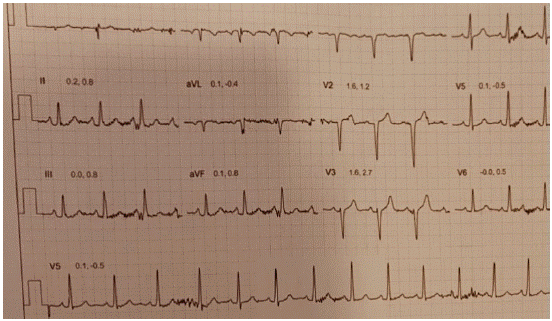
Figure 1:
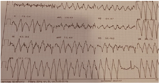
Figure 2:
Referral was made for an urgent outpatient angiogram to assess for coronary artery disease and repeat echocardiography was performed to assess for structural heart disease which showed a normal ejection fraction and normal valvular function with no foreign bodies.
Angiography did not show any significant coronary artery disease however a small metallic artefact was seen in the ventricle, (Figures 3 & 4). With patient consent further angiography of the right forearm at the site of previous shrapnel injury was performed and no metallic artefact was identified.
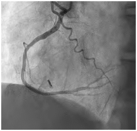
Figure 3:
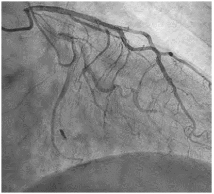
Figure 4:
Cardiac CT imaging was performed which identified a 5 mm intracardiac metallic structure within the right ventricle (Figure 6).
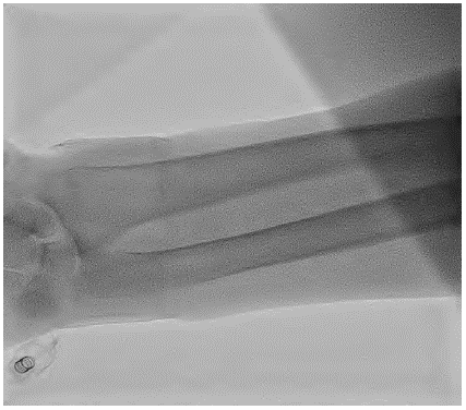
Figure 5:
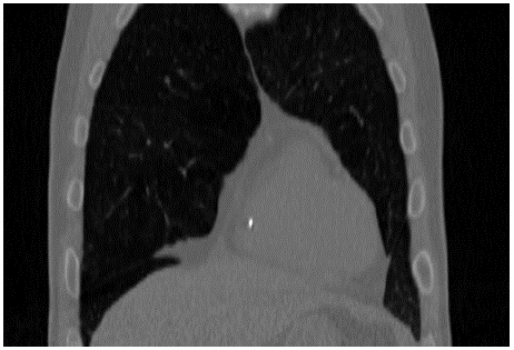
Figure 6:
Given the patients co-morbidities and a subsequent diagnosis of lung malignancy on the Coronary CT, conservative management was adopted, and he was started on low dose Bisoprolol. He underwent lobar lung resection for a bronchogenic neoplasm without intervention on the metallic artefact after multi-disciplinary discussion. He had clear margins and fifteen negative lymph nodes at the time of surgery. The patient remained well with no symptomatic arrhythmias throughout the periop and post op period and recovered well.
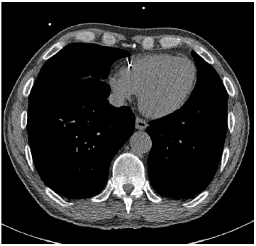
Figure 7:
Given the patient’s co-morbidities and a subsequent diagnosis of lung malignancy on the Coronary CT, conservative management was adopted for the metallic device and he was started on low dose bisoprolol. He underwent lobar lung resection for a bronchogenic neoplasm without cardiac intervention on the metallic artefact. He had clear margins and fifteen negative lymph nodes at the time of surgery.
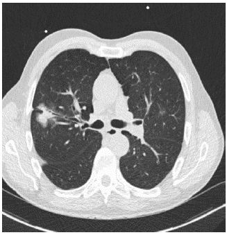
Figure 8:
The patient remained well with no symptomatic arrhythmias throughout the operative period and recovered well. Cardiac follow up six months post operatively did not demonstrate any new cardiac symptoms, palpitations, syncopal episodes or chest pains. There was no clinical feature of local or metastatic spread at six months of surgical follow-up.
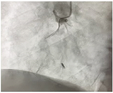
Figure 9:
Cardiac foreign bodies present with a wide range of mechanisms from penetrating trauma, iatrogenic injuries, malignant dysrhythmias, accidents and from venous emboli from other sites, predominantly to the right side of the heart [3,4]. They remain difficult to treat and clear evidence-based guidelines on diagnosis and management is lacking. Leitman et al analysed 104 cases of patients presenting with cardiac foreign bodies. They concluded Iatrogenic emboli as the predominant cause followed by accidents and missiles. Of the 14 IVC filter related cases three initially presented with cardiorespiratory arrest, highlighting a need for prompt diagnosis [5]. However, these cases are difficult to diagnosis and a high index of suspicion is often needed. In this case, diagnosis of both the foreign body and the subsequent lung cancer were incidentally found. With regards to diagnosis, chest x-ray may be useful for identifying opaque foreign bodies while echocardiography is recommended for localising the foreign body and assessing its movement during and interaction with the cardiac cycle [6]. In this case both transthoracic echo and chest x-ray failed to identify the metallic object and it was only with the use of exercise stress testing, coronary angiography and CT that the object was characterised. Without the intracardiac foreign body the resulting lung malignancy would not have been identified.
Management guidance in the literature is scarce, one systematic review assessing management of cardiac foreign bodies from sewing injuries favoured surgical intervention over conservative treatment even in asymptomatic patients [7]. It is hypothesised that sharper and pericardial foreign bodies are of more danger with higher rates of pericarditis and pericardial effusions, while those that are endocardial and present for longer durations may become embedded in the heart and thus be less clinically significant [8-10]. Surgical treatment is also dependent on the type of foreign body and the need for cardiopulmonary bypass [9]. Furthermore there have also been successful retrievals of iatrogenic foreign bodies using intravascular catheters [11]. In this case, medical co-morbidities and the subsequent diagnosis of a lung cancer on CT favoured a conservative approach to management of the foreign body.
Learning Points
- Foreign bodies are a potential cause of ventricular ar rythmias.
- Identification of foreign bodies in the ventricle on an giography.
- Management and challenges of cardiac foreign bodies.
- Imaging review.
• Review of published literature on diagnosis and management of cardiac foreign bodies.
References
- Wang X, Zhao X, Du D, Xiang X. Management of Metallic Foreign Bodies in the Heart. Journal of Cardiac Surgery. 2012; 27: 704-6.
- Symbas PN, Picone AL, Hatcher CR, Vlasis-Hale SE. Cardiac missiles. A review of the literature and personal experience. Ann Surg. 1990; 211: 639-48.
- Baker CJ, Nigro JJ, Daggett CW, Wells WJ. Needle embolism to the heart. The Annals of thoracic surgery. 2004; 77: 1102.
- Willemsen P, Kuo J, Azzu A. Dysrhythymia from an intrapericardial air gun pellet: a case report. Eur J Cardio-thorac Surg. 1996; 11996: 461-2.
- Leitman M, Vered Z. Foreign Bodies in the Heart. Echocardiography. 2015; 32: 365-71.
- Fry SJ, Picard MH, Tseng JF, Briggs SM, Isselbacher EM. The Echocardiographic Diagnosis, Characterization, and Extraction Guidance of Cardiac Foreign Bodies. Journal of the American Society of Echocardiography. 2000; 13: 232-9.
- Perrotta S, Perrotta A, Lentini S. In patients with cardiac injuries caused by sewing needles is the surgical approach the recommended treatment? Interactive cardiovascular and thoracic surgery. 2010;10 5:783-92.
- LeMaire SA, Wall Jr MJ, Mattox KL. Needle embolus causing cardiac puncture and chronic constrictive pericarditis. The Annals of thoracic surgery. 1998; 65: 1786-7.
- Actis Dato GM, Arslanian A, Di Marzio P, Filosso PL, Ruffini E. Posttraumatic and iatrogenic foreign bodies in the heart: report of fourteen cases and review of the literature. J Thorac Cardiovasc Surg. 2003; 126: 408-14.
- Perrotta S, Perrotta A, Lentini S. In patients with cardiac injuries caused by sewing needles is the surgical approach the recommended treatment? Interactive Cardio Vascular and Thoracic Surgery. 2010; 10: 783-92.
- Khaja F, Lakier J. Foreign body retrieval from the heart by two catheter technique. Catheterization and cardiovascular diagnosis. 1979; 5: 263-8.