Case Report
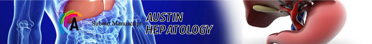
A 61 years old men with no past medical facts, presented in the emergency for acute obstructive dysphagia. Upper endoscopy showed an impacted foreign body (chicken bone) impacted in the middle oesophagus which was removed successfully. Control endoscopy identified a bulbar ulcerated polylobed submucosal tumor measuring 2-3 centimeters. Echoendoscopic ultrasound found a heterogenous hypoechogenic tumor developing from the 3-4th couch (Figure 1-2). CT scan found rregular non-stenotic thickening of the bulbar wall with no other localizations (Figure 3). Biopsy of the tumor showed a mucosal proliferation of monomorophous cells, forming nests in vascular stroma with positivity of synaptophysin in immunostaining suggestive of neuroendocrine tumor (Figure 4). Patient underwent enucleation successfully (Figure 5).
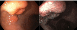
    Figure 1: Upper endoscopy showing a submucosal polylobate ulcerated
tumor of 2-3 centimeters.
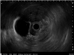
    Figure 2: Echo endoscopic ultrasound: Heterogeneous hypo echogenic
tumor developing from the 3-4th couch measuring 5×3 centimeters.
    Figure 3: CT scan objectifying irregular non-stenotic thickening of the bulbar
wall.
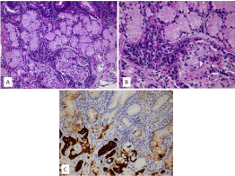
    Figure 4: Infiltration of the bulb AR mucosa by a neuroendocrine tumor.
A: mucosal proliferation of neuroendocrine cells (H&E staining × 200)
B: tumor cells are monomorphous, forming nests in vascular stroma (H&E
staining × 400)
C: Immunostaining for Synaptophysin (× 200)
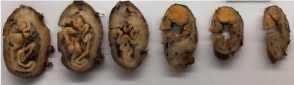
Figure 5: Macroscopic aspect of the enucleation of the lesion.