
Abstract
Hypercalcemia can be a manifestation of malignancy after other causes like hyperparathyroidism are ruled out. We describe a case of Marjolin’s ulcer in a young female, which was diagnosed after the initial manifestation of hypercalcemia. 28-year-old female with history of paraplegia, chronic decubitus ulcers, who came into the Emergency department with fever, vomiting and pain at the ulcer site. She was noted to have an ulcer in the right ischial tuberosity. Labs revealed hypercalcemia. She was admitted for acute on chronic osteomyelitis and started on intravenous antibiotics. Calcium levels remained persistently elevated despite hydration and calcitonin administration. Malignancy was suspected and she underwent punch biopsies from the ulcer. Pathology showed moderately differentiated squamous cell carcinoma. Parathyroid Hormone-Related Protein (PTH-rp) was slightly elevated. CT abdomen pelvis revealed large right inguinal and pelvic lymph nodes. CT thorax revealed a right lower lobe mass, with two additional nodules in the right upper lobe and associated hilar lymphadenopathy. She was given zoledronic acid to treat the hypercalcemia to which she responded. Humoral hypercalcemia of malignancy is secondary to the production of humoral factors by malignant cells in patients without bony metastases. PTH-rp is the main factor implicated. Case reports have also mentioned cytokines and transforming growth factor alpha production as a cause of hypercalcemia in patients with Marjolin’s ulcer. Hypercalcemia should raise suspicion for malignant transformation in a patient with chronic decubitus ulcers, despite the age of the patient and the duration of ulcer.
Keywords: Hypercalcemia; Marjolin’s ulcer; PTH-rp; Squamous cell
Abbreviations
HHM: Humoral Hypercalcemia of Malignancy; PTH-rp: Parathyroid Hormone-Related Protein
Introduction
Unexplained hypercalcemia can be a manifestation of malignancy after other causes like hyperparathyroidism are ruled out. Humoral Hypercalcemia of Malignancy (HHM) is a paraneoplastic syndrome that can occur in a wide variety of cancers. This is secondary to the production of humoral factors by malignant cells in patients without bony metastases [1]. Case reports have described the occurrence of hypercalcemia in squamous cell carcinoma of the skin [2-5]. Marjolin’s ulcer is the malignant transformation of long-standing ulcers, burns and fissures. Hypercalcemia can sometimes be the only initial clue to an occult malignancy. We describe a case of Marjolin’s ulcer in a young female, which was diagnosed after the initial manifestation of hypercalcemia.
Case Presentation
28-year-old female with history of paraplegia since 9 years of age following a sledding accident, chronic decubitus ulcers with osteomyelitis, status post diverting colostomy, who came into the Emergency Department with fever, chills, malaise, nausea, vomiting and pain at the site of her ulcers for the past 5 days. On exam, vitals were stable. She was noted to have 15x15x15 cm ulcer in the right ischial tuberosity with exposed deep tissues, palpable bone with a fibrinous foul smelling exudate at the base and an additional ulcer in the right greater trochanter tracking to the ischial ulcer along with erosion of the right labial fold. She also had bilateral lower extremity weakness with sensory loss. Labs revealed WBC of 10.9x10³/ul with neutrophilic predominance, microcytic anemia with hemoglobin of 10.9 g/dl and hematocrit of 34%, platelets of 667x103/ul, ESR of 47 mm/hr, CRP of 113 mg/l, sodium of 133 mmol/l, potassium of 4.3 mmol/l, chloride of 94 mmol/l, bicarbonate of 27 mmol/l, blood urea nitrogen of 5 mg/dl and creatinine of 0.4 mg/dl. She was also noted to have elevated calcium levels of 14 mg/dl (corrected value of 14.5 mg/ dl) and ionized calcium of 1.97 mmol/l. She was admitted for acute on chronic osteomyelitis and started on piperacillin/tazobactam along with intravenous hydration for hypercalcemia. Endocrine consult was done. Work up for hypercalcemia revealed normal alkaline phosphatase of 106 U/l, 25-hydroxy Vitamin D of 8 ng/ml and parathyroid hormone of 3 pg/ml. Parathyroid Hormone-Related Protein (PTH-rp) was sent. Serum protein electrophoresis showed hypoalbuminemia with elevated acute phase reactants. Calcium levels remained persistently elevated despite intravenous hydration and calcitonin administration. With the intractable hypercalcemia, malignancy was suspected and she underwent multiple punch biopsies from the decubitus ulcer. Pathology showed moderately differentiated squamous cell carcinoma positive for high-risk human papilloma virus (Figure 1,2 and 3). PTH-rp came back slightly elevated at 28 pg/ml (Reference range of 14 - 27 pg/ml). CT abdomen pelvis with contrast showed extensive soft tissue ulceration with gas formation and destruction of a large portion of the right hip and hemi pelvis along with large right inguinal and pelvic lymphadenopathy, concerning for metastatic disease (Figure 4). CT thorax with contrast revealed a right lower lobe mass of 3 cm, with two additional nodules in the right upper lobe of 1.5 cm and 4 mm along with associated right hilar lymphadenopathy also suggestive of metastasis. Multiple imaging studies failed to reveal any metastatic bone lesions. She was given zoledronic acid to treat the hypercalcemia to which she responded. She also underwent CT guided fine needle aspiration of the lung mass, which also revealed squamous cell carcinoma. She was later discharged with oncology follow up for further management.
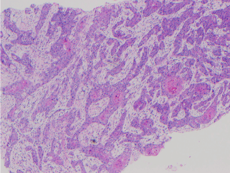
    Figure 1: Hematoxylin and eosin stain of the perineal lesion showing
moderately differentiated squamous cell carcinoma.
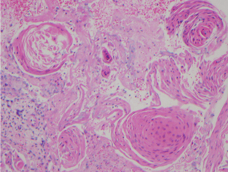
Figure 2: Shows abundant keratinization within the tumor.
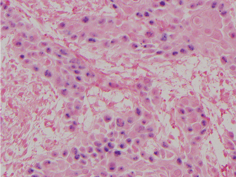
    Figure 3: High power showing Human Papilloma Virus in situ hybridization
positivity.
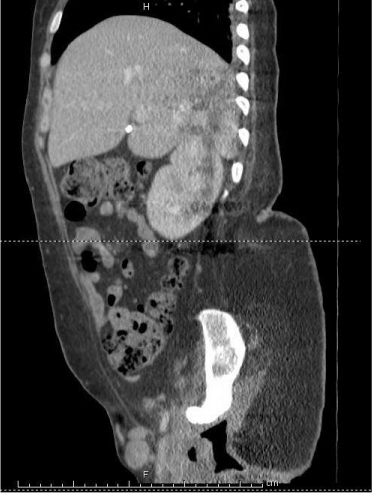
    Figure 4: CT abdomen pelvis with contrast showed extensive soft tissue
ulceration and large right inguinal lymphadenopathy.
Discussion
Hypercalcemia is a relatively common problem encountered in clinical practice. Among all the causes of hypercalcemia, primary hyperparathyroidism and malignancy accounts for greater than 90 percent of the cases and is secondary to bone resorption. Although primary hyperparathyroidism can occur at any age, calcium levels greater than 13 mg/dl is typically rare. Mild hypercalcemia can be seen in around 15 to 20 percent of patients with thyrotoxicosis secondary to the above mechanism [6]. Other causes like prolonged immobilization, Paget’s disease and hypervitaminosis A increases bone resorption and causes hypercalcemia. Hypervitaminosis D, that can be seen in increased dietary intake of Vitamin D or increased endogenous production of 1, 25-dihydroxyvitamin D in granulomatous diseases cause hypercalcemia by increased calcium absorption and bone resorption [7]. Other causes of hypercalcemia include milk alkali syndrome, adrenal insufficiency, pheochromocytoma and drugs such as lithium, thiazide diuretics and theophylline. Familial hypocalciuric hypercalcemia and metaphyseal chondrodysplasia are rare causes.
Hypercalcemia occurs in approximately 20 to 30 percent of patients with cancers [8]. While osteolytic metastasis is the cause in 20% of the patients, HHM accounts for 80% of the patients. PTH-rp is the main factor implicated in HHM. It stimulates reabsorption of calcium in the distal collecting ducts of kidney and causes osteoclastic bone resorption by interacting with PTH receptors [9]. HHM can also be secondary to the ectopic secretion of PTH or 1, 25-dihydroxyvitamin D [10]. Tumor necrosis factor-beta has been implicated as a factor in adult T-cell leukemia-lymphoma [11]. In squamous cell carcinomas, epidermal growth factor and transforming growth factor-alpha have been implicated as stimulators of bone resorption and hypercalcemia [12,13,14]. As PTH-rp was only marginally elevated in our patient, HHM could have been secondary to any of the above mechanisms. On literature review, there have been only around 4 case reports of hypercalcemia described in Marjolin’s ulcer [4,5,15,16]. Other possible mechanisms of hypercalcemia to be considered in this patient are prolonged immobilization, [15,16], which is unlikely as she had been paraplegic for a long time prior to presentation and possible primary hyperparathyroidism, which was ruled out.
Marjolin’s ulcers are relatively rare, seen in around 0.7% of scars as noted in a study [17]. The time necessary for malignant transformation ranges from 20 to 70 years, averaging around 30 years [2,18]. However, our patient was younger than the average age mentioned above which makes initial suspicion of malignancy unusual. In squamous cell carcinomas, it has been noted that tumor excision has been shown to improve hypercalcemia [19]. This was a difficult option in our patient given her extensive ulcers.
Marjolin’s ulcers usually have an aggressive course and associated with a poor prognosis [20]. HHM usually has a poor prognosis, but the prognosis with HHM associated with Marjolin’s ulcer is less ominous [15].
Conclusion
Resistant hypercalcemia should raise suspicion for malignant transformation in a patient with chronic decubitus ulcers despite the young age of the patient and duration of the ulcer. Early diagnosis and management can possibly impact survival in these patients.
References
- Szymanski JJ, Otrock ZK, Patel KK, Scott MG. Incidence of humoral hypercalcemia of malignancy among hypercalcemic patients with cancer. Clinica Chimica Acta. 2016; 453: 190-193
- Reynaud-Mendel B, Robert C, Flageul B, de Vernejoul MC, Verola O, Dubertret L. Malignant hypercalcemia induced by a parathyroid hormonerelated protein secreted by a cutaneous squamous cell carcinoma. Archives of dermatology. 1997; 133: 113
- Cisneros G, Lara LF, Crock R, Whittier FC. Humoral hypercalcemia of malignancy in squamous cell carcinoma of the skin: parathyroid hormone- -related protein as a cause. Southern medical journal. 2001; 94: 329-331.
- O’Malley JT, Schoppe C, Husain S, Grossman ME. Squamous Cell Carcinoma (Marjolin’s Ulcer) Arising in a Sacral Decubitus Ulcer Resulting in Humoral Hypercalcemia of Malignancy. Case reports in medicine. 2014; 13.
- Hall AB, Buehler KE, Philipneri M. Hypercalcemia Complicating Marjolin’s Ulcer. Military medicine. 2009; 174: 308-310.
- Hall AB, Buehler KE, Philipneri M. Hypercalcemia Complicating Marjolin’s Ulcer. Military medicine. 2009; 174: 308-310.
- Jacobs TP, Bilezikian JP. Rare causes of hypercalcemia. The Journal of Clinical Endocrinology & Metabolism. 2005; 90: 6316-6322.
- Stewart AF. Hypercalcemia associated with cancer. New England Journal of Medicine. 2005; 352: 373-379.
- Sparks MK, Kuhlman DS, Prieto A, Callen JP. Hypercalcemia in association with cutaneous squamous cell carcinoma: occurrence as a late complication of hidradenitis suppurativa. Archives of dermatology. 1985; 121: 243-246.
- Horwitz MJ, Stewart AF. Hypercalcemia associated with malignancy. Primer on the metabolic bone diseases and disorders of mineral metabolism. 2006; 6: 195-199.
- Ishibashi K, Ishitsuka K, Chuman Y, Otsuka M, Kuwazuru Y, Iwahashi M, et al. Tumor necrosis factor-beta in the serum of adult T-cell leukemia with hypercalcemia. Blood. 1991; 77: 2451-2455.
- Ibbotson KJ, D’souza SM, Ng KW, Osborne CK, Niall M, Martin TJ, et al. Tumor-derived growth factor increases bone resorption in a tumor associated with humoral hypercalcemia of malignancy. Science. 1983; 221: 1292-1294.
- Raisz LG, Simmons HA, Sandberg AL, Canalis E. lDirect Stimulation of Bone Resorption by Epidermal Growth Factor*. Endocrinology. 1980; 107: 270- 273.
- Mundy GR. Pathophysiology of cancer-associated hypercalcemia. In Seminars in oncology. 1990; 17: 10-15.
- Sungur N, Kilinc H, Uysal C, Ortak T, Sensoz O. A gigantic squamous cell carcinoma with hypercalcemia arising in an old burn scar. Annals of plastic surgery. 2001; 47: 351-352.
- Gerner RE, Moore GE. Burn scar carcinoma and hypercalcemia. Annals of surgery. 1974; 180: 95.
- Yu N, Long X, Lujan-Hernandez JR, Hassan KZ, Bai M, Wang Y, et al. Marjolin’s ulcer: a preventable malignancy arising from scars. World journal of surgical oncology. 2013; 11: 1.
- Kerr-Valentic MA, Samimi K, Rohlen BH, Agarwal JP, Rockwell WB. Marjolin’s ulcer: modern analysis of an ancient problem. Plastic and reconstructive surgery. 2009; 123: 184-191.
- Loche F, Bennet A, Bazex J, Thouvenin MD. Hypercalcaemia of malignancy associated with invasive cutaneous squamous cell carcinoma. The British journal of dermatology. 1999; 141: 577-579.
- Tobin C, Sanger JR. Marjolin’s Ulcers: A Case Series and Literature Review. Wounds: a compendium of clinical research and practice. 2014; 26: 248-254.