
Introduction
Congenital enlargement of extraocular muscles is an uncommon finding. Few cases have been published. Dickson JS, et al. [1], reported in 1994 the findings of an 8-months-old girl with unilateral left esotropia and hypotropia. The orbit Computer Tomography (CT) scan showed enlargement of left inferior, lateral and medial rectus muscles. No abnormality was found in studies of a biopsy specimen. Later in 2006, Burroughs JR et al. [2] reported a 2-months-old boy presented with left eye hypotropia, exotropia and proptosis at birth. Orbit CT scan showed enlarged extraocular muscles. Muscle biopsy and thyroid profile were normal. More recently, Kekunnaya R, et al [3], reported the case of a 20-months-old boy with non-progressive downward deviation of the right eye since 10 months of age. A CT scan of the orbits showed diffuse thickening of the posterior part of the right inferior rectus. Thyroid profile was normal and muscle biopsy showed non-specific fibrotic changes. In this report, we present the clinical findings of one patient with unilateral congenital enlarged extraocular muscles.
Case Presentation
A 4-month-old boy was referred with history of left eye hypotropia at birth. The patient was otherwise healthy, no history of prematurity, normal gestation and birth. There was no history of systemic affection of the mother. The patient had central fixation with both eyes, no nystagmus. Epiblefaron was observed and he had clear media with a normal anterior segment in both eyes. No proptosis was documented. A left exotropia and hypotropia were found (Figure 1A). Ocular movements were full in the right eye. A marked limitation to elevation of the left eye was noted. Fundus exam was normal in both eyes. The orbit CT scan showed enlarged inferior and lateral rectus muscle in left eye (Figure 1B). Forced duction test showed remarkable restriction to elevation of the left eye. Hematology and thyroid profile were normal. The family of the patient abandoned the follow-up. Planning of strabismus surgery was not possible.
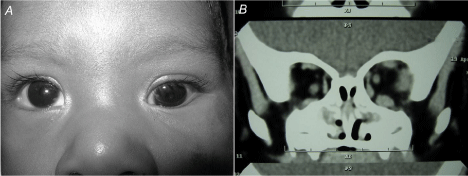
Figure 1A & 1B: A left exotropia and hypotropia.
Discussion
The differential diagnosis in extraocular muscle enlargement includes infiltrative, inflammatory, neoplastic and vascular processes [2]. In the pediatric population finding congenital extraocular muscle enlargement is uncommon, with little information regarding etiology and presentation. Although the cases reported in the literature may give some ideas, this entity still represents a non-diagnostic congenitally and unilaterally enlargement of extra ocular muscle. It may be related to a congenital thyroid eye disease in euthyroid patient and mother, or part of congenital unilateral fibrosis, blepharoptosis and enophthalmos syndrome [1-3]. In our differential diagnosis we included thyroid eye disease, but unlike congenital hypothyroidism, neonatal hyperthyroidism is a rare entity, almost always transient and is usually caused by transplacental transmission of antithyroid antibody from a dysthyroid mother [4]. In our case the thyroid profile was normal. Other diagnosis to be considered is congenital orbital fibrosis [5]. Our case presented forced ductions test with restriction on left eye, but did not have enophthalmos which is more characteristic of this diagnosis.
As a summary, constant findings in all reported cases are: male gender, hypotropia and exotropia, supraduction inability, enlarged inferior and lateral rectus muscles, forced duction test with restriction and normal thyroid profile. Proptosis, enophthalmos and pseudoptosis are inconstant. We do not have histology studies in our case, but the results in the other reports are not conclusive [1-3]. These cases follow a pattern, and the many common findings support the idea that we could be dealing with the same entity, waiting to be fully discovered. More studies to find answers will be needed.
References
- Dickson JS, Kraft SP, Jay V, Blaser S. A case of unilateral congenitally enlarged extraocular muscles. Ophthalmology. 1994; 101: 1902-1907.
- Burroughs JR, Bearden WH, Anderson RL, Hoffman RO, Elliot RL, Mccann JD. Congenitally enlarged extraocular muscles: can congenital thyroid eye disease exist in a euthyroid infant? ophthal plast reconstr surg. 2006; 22: 314-316.
- Kekunnaya R, Bansal R, Vemuganti GK. Congenitally dysplastic inferior rectus muscle. J Pediatr Ophthalmol Strabismus. 2010; 22: 47.
- Brown RS. Neonatal Hyperthyroidism. In: De Groot LJ, Beck-Peccoz P, Chrousos G, Dungan K, Grossman A, Hershman JM, et al. Endotext [Internet]. South Dartmouth (MA): MDText.com, Inc. 2000.
- Li YJ, Han J, Yan H, Li J, Wang D, Xu S. Congenital orbital fibrosis associated with fibrosis of extraocular muscle. BMJ case reports. 2012; 10.