
Clinical Image
J Bacteriol Mycol. 2016; 3(1): 1023.
Neurobrucellosis in a Striped Dolphin
Di Guardo G*
Faculty of Veterinary Medicine, University of Teramo, Località Piano d’Accio, 64100 - Teramo, Italy
*Corresponding author: Di Guardo G, Faculty of Veterinary Medicine, University of Teramo, Italy
Received: April 04, 2016; Accepted: April 20, 2016; Published: April 22, 2016
Clinical Image
The image shows a brain tissue section obtained from a striped dolphin (Stenella coeruleoalba) (Figure 1), which was found stranded in 2012 along the southern Adriatic Sea coast of Italy. A prominent, subacute-to-chronic, predominantly lympho-histiocytic leptomeningitis is observed, along with a simultaneous, though far less pronounced involvement of the subcortical grey matter by a similar inflammatory infiltrate. The agent responsible for the aforementioned lesions’ development is Brucella ceti, the most recent “entry” into the Brucella genus, a feature which is also shared by B. pinnipedialis. Besides infecting a consistent number of aquatic mammal species worldwide, B. ceti and B. pinnipedialis are zoonotic pathogens, capable of causing neurobrucellosis and osteomyelitis in human patients. Likewise, B. ceti is known to display a strong neurotropism in striped dolphins, which frequently develop remarkable brain lesions particularly affecting their meninges (as in the case presented here) and choroid plexuses, with subsequent neurological impairment, disorientation and stranding.
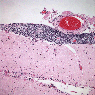
    Figure 1: Brain tissue section obtained from a striped dolphin (Stenella
coeruleoalba).