
Case Report
Austin Biomark Diagn. 2015; 2(2): 1022.
Giant Chondrosarcoma in Ollier’s Disease
Zvonimir Zore* and Mladen Stanec
Department of Surgical Oncology, University Hospital Center Sestre milosrdnice, Zagreb, Croatia
*Corresponding author: Zvonimir Zore, Department of Surgical Oncology, University Hospital Center Sestre milosrdnice, Zagreb, Croatia
Received: July 08, 2015; Accepted: November 24, 2015; Published: November 26, 2015
Keywords
Ollier’s disease; Enchondromatosis; Chondrosarcoma
Summary
A 30-year-old male presented with increasing tumors size, limited motion and pain in the left upper arm of approximately 5 years duration, surgically treated 15 years ago. Final histopathological exam was confirmed clinically diagnosis, that it was well-differentiated giant chondrosarcoma. The patient is alive and well 15 years after amputation upper limb.
Introduction
Enchondromas (Multiple Enchondromatosis, Dyschondroplasia, and Enchondromatosis) are common intraosseous benign cartilaginous tumors. When multiple enchondromas are present, the condition is called enchondromatosis, also known as an Ollier’s disease by the terminology of the World Health Organization [1]. Ollier’s disease is very rare. The estimated prevalence of Ollier’s disease is 1/100,000 [2]. Boys are affected twice as often as girls. The disease tends to be bilateral but predominate on one side and primarily affects the long bones and cartilage of the joints of the arms and legs. It may occur at anytime. Symptoms may also arise secondary to mass effect resulting in pain and loss of motion, compression or stretching peripheral nerves and pathological fractures. The diagnosis is based on clinical and conventional radiological evaluations. Surgery is indicated in case of complications: pathological fractures, growth defect and malignant transformation. The reported incidence of malignant trasformation (e.g., chondrosarcomas) is variable and estimated to occur in 5-50% [3].
Chondrosarcoma-Case Report
In 1998, a 30 year old male patient was presented with a dominant tumorous mass of the proximal left humerus (Figure 1). He had been operated few ways, last operation was performed for 10 years previously because he had multiple exostosis since childhood. The patient has had not completed documentation for previously operations, only for last operation. That operation has been done on both forearms because there was exostosis. After that patient has had fractures of both forearms. Physical exam showed no abnormalities except a tumorous mass of proximal left humerus sized approximately 30 cm, reduced length of both forearms and exostosis on both proximal lower legs.Patient has had two times a transient peripheral paralysis of nerves facials. Intraoral findings are massing on the hard palate look like torus palatinus (exostosis on the hard palate). In patients family nobody has the same mass. Plain radiograph shows a large ill-defined, destructive, humerus metaphysis intramedullary lesion with bone destruction and periosteal reaction (Figure 2). The lesion is associated with a soft tissue mass. Furthermore, plain radiographs showed hiperostotic change of the lateral part of right clavicle, metaphysis of neck and trochanter of both femurs, bones of head and intraoral findings. Scintigraphy showed enhanced accumulation radionuclide on proximal right humerus, right shoulder and right trochanter of femur. CT demonstrated chondrosis of metaphysis of right humerus and cranial part of the shoulder blade. Preoperative biopsies showed chondrosarcoma.
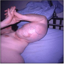
Figure 1: Clinical findings.
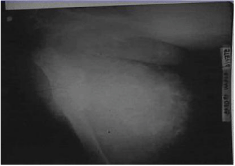
Figure 2: Characteristic radiological findings.
Operation
After incision of skin from acromio-clavicular joint to lateral epicondyle, the fascia had been dissected where a 30cm tumor had been found. It had a very good blood supply. All of tumorous mass was showed and dissecting from n. radialis. Due to operations distal part of the humerus was intact, but at the medial and proximal part of humerus junction corticalis was damaged with extension of the tumor into soft tissues. There was bone medulla fulfilled with gelatinous material. We canceled of tumorectomy because of the above mentioned conditions and indicated disarticulation in the shoulder joint.
The Final histopathological exam was confirmed clinically diagnosis that it was good differentiated chondrosarcoma. We performed a head CT because there was suspected paralysis of the facial nerve and possibly of intracranial metastasis. There were no suspect findings, but transient facial nerve paralysis was probably resulted of hyperossification of petrous bone and consequent nerve ischemia. 15 years after operations classical radiological and scintigraphic findings does not show suspect metastasis or received. The patient is alive and well 15 years after operative treatment.
Discussion
Malignant transformation (mutation) in enchondromatosis is well recognized and is the most common neoplasm occurring in patients with multiple enchondromas. Malignant transformation can occur during childhood or adolescence, but the risk increases with age [3]. The difficult diagnostic dilemma is distinguishing enchondroma from low-grade chondrosarcoma. Pain in enchondroma is a worrisome symptom, which indicates either a pathologic fracture or continued growth. The large size of the lesion > 5cm, certain skeletal locations (it is more common in long bones than short) and “aggressive” radiological features of the lesion favor the diagnosis of chondrosarcoma [4]. The malignant transformation is usually a slow process, occurring over decades. Since Ollier’s disease is self-limited in that it usually stops spontaneously as the patient grows, and since the cartilaginous lesions in occasional cases may regress or even disappear, any cartilaginous lesions that are still active or painful after termination of the growth period should be examined thoroughly under suspicion of undergoing malignant transformation [4,5]. When enchondromas are found in the long bones or axial skeleton, there is a higher risk (44%-50%) for developing chondrosarcomas. The mean age at which the patients first underwent surgery for chondrosarcoma was 33 years for patients with Ollier disease [6].
Signs of malignant transformation in the presented patient include metaphyseal location, long bones, the large size of the tumor (>10cm), cortical erosion, extension of the tumor into soft tissues, irregularity of the surface of the tumor, a long duration of symptoms that can be associated with worse prognosis of the patient. Younger age, low histological grade, local surgical stage and radical resections have been associated with better prognosis and longer survival. The prognosis for Ollier’s disease in the reported patient is difficult to assess. Chondrocasrcomas grade I can recur after 10 to 20 years [7,8]. Patients with malignant transformation from enchondroma towards grade I chondrosarcoma have a 5 year survival rate of 90%, respectively, and 10 year survival rate of 83% [7,8].
References
- Slive C, Juppner H. Ollier disease. Orphanet J Rare Dis. 2006; 1: 37.
- Giuffrida AY, Burgueno JE, Koniaris LG, Gutierrez JC, Duncan R, Scully SP. Chondrosarcoma in the United States (1973 to 2003): an analysis of 2890 cases from the SEER database. J Bone Joint Surg Am. 2009; 91: 1063-1072.
- Pannier S, Legeai-Mallet L. Hereditary multiple exostoses and enchondromatosis. Best Pract Res Clin Rheumatol. 2008; 22: 45-51
- Mavrogenis AF, Gambarotti M, Angelini A, Palmerini E, Staals EL, Ruggieri P, et al. Chondrosarcomas Revisited. Orthopedics. 2012; 35: 379- 390.
- Verdegaal SH, Bovée JV, Pansuriya TC, Grimer RJ, Ozger H, Jutte PC, et al. Incidence, predictive factors, and prognosis of chondrosarcoma in patients with Ollier disease and Maffucci syndrome: An International Multicenter Study of 161 patients. Oncologist. 2011; 16: 1771-1779.
- Vazquez-Garcia B, Valverde M, San-Julian M. Ollier disease: benign tumours with risk of malignant transformation. A review of 17 cases. An Pediatr. 2011; 74: 168-173.
- Herget GW, Strohm P, Rottenburger C, Kontny U, Krauß T, Bohm J, et al. Insights into Enchondroma, Enchondromatosis and the risk of secondary Chondrosarcoma. Review of the literature with an emphasis on the clinical behaviour, radiology, malignant transformation and the follow up. Neoplasma. 2014; 61: 365-378.
- Giuffrida AY, Burgueno JE, Koniaris LG, Gutierrez JC, Duncan R, Scully SP. Chondrosarcoma in the United States (1973 to 2003): an analysis of 2890 cases from the SEER database. J Bone Joint Surg Am. 2009; 91:1063-1072.