
Research Article
Austin J Biotechnol Bioeng. 2017; 4(1): 1070.
Assessment of Silver Nanoparticle Formation from Non- Pathogenic Enteric Bacteria using SEM
Saklani V*
Amity University, India
*Corresponding author: V, Amity University, Noida, U.P, India
Received: November 21, 2016; Accepted: March 09, 2017; Published: March 16, 2017
Abstract
The following paper proposes the analysis of silver nanoparticle formation using SEM (Scanning Electron Microscopy). The analysis is based on the information through experiments conducted in the laboratory. It provides a clear picture of the nanoparticle formation in terms of temperature, color change, concentration of mixture, agglomeration and finally visualizing the effects through SEM. The study also consists of the comparison of toxicity of silver nanoparticle against E. coli, at three different concentrations; 0.1mM, 1mM, 10mM of silver nitrate at 10% v/v mixture.
Keywords: Scanning electron microscopy; Crystallization; Silver nanoparticle; E. coli; Biomineralisation
Abbreviations
SEM: Scanning Electron Microscopy; Crystallization; Silver Nanoparticle; E. coli: Escherichia coli; Biomineralisation
Introduction
The production of nanoparticles from microbes is a boom in the field of material sciences. Many scientists from all over the world are using microbes for the generation of nanoparticles. The study consists of the generation of silver nanoparticles from an enteric bacterium, E. coli. It is a non-pathogenic and can easily be retrieved. In this paper, we reported the mechanism underlying the generation of silver nanoparticles from E. coli and with a concentration of 10% v/v mixture.
The nanoparticles are formed from the inoculated nutrient broth. This is a very interesting topic in the field of biomineralisation and forms the basis of this study. Usually experiments conducted at invitro level made use of silver nitrate of 1mM at 1% v/v concentration of mixture (was considered the standard concentration) to produce nonmaterial’s. The mechanism lies in the fact that several factors are responsible for the formation of crystals.
The concentration of silver nitrate is very less but the microbes are very active, they are able to make crystals very fast. For this we studied the crystal formation at two different temperature conditions. One at 37 degrees Celsius and other at Room Temperature (RT). E. coli, non-pathogenic strain forms a part of normal flora of the human gut. The optimum temperature for bacterial growth is 37 degrees Celsius. It is usually said that microbial action is slow in experimentation [1- 19]. But the production of nanoparticles here was fast as silver nitrate acts as an external trigger for the bacterial machinery and thus they were able to quickly generate nanoparticles. The mechanism behind says that according to the microbial resistance to toxic silver ions in the environment, the bacterial machinery is adapted to convert these toxic ions into stable metal, i.e. silver metal. This is a result of biomineralisation. Hence, we can also say that E. coli is the production house for silver metal. If we look towards the pH side, normally E. coli in the gut flora is slightly alkaline, i.e. 6-7. In the inoculated NB (Nutrient Broth), the pH was 6.89.
The formation of crystals depends on the bacterial metabolism and the outer environment of the bacteria where it is residing. According to the analysis, as studied by other scientists as well, the UV-Visible graph showed the slight shift in the peaks and the absorption was in both the UV and visible region. This indicated the plasmon effect of the silver nanoparticles. The variation in the peak shift with time showed the nanoparticle formation. However, the toxicity of silver nanoparticle varies with the concentration of silver nitrate [1].
There was also an encapsulation of nanoparticles in the biofilm and thus can be said that E. coli acts as a natural generator of nanoparticles and the concentration of silver nitrate can help it in the controlled release of metal nanoparticles in the surrounding. It can be said that E. coli served as a reservoir of silver at 0.1mM AgNO3 concentration (Figures 1-3). As the concentration went higher (i.e. 10mM) there was an accumulation of silver nanoparticles leading to cell death (Figures 4 &5).
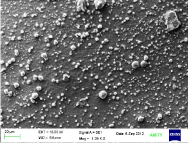
    Figure 1: The figure demonstrates the silver nanoparticles formed by the
bacterial enzymatic reduction analyzed through SEM.
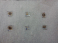
    Figure 2: The picture shows the glass pieces on which the sample is mounted
for SEM stage. They are at three different concentrations and at two different
temperatures.
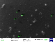
Figure 3: The silver nanoparticle formation at 0.1mM (AgNO3) concentration.
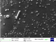
Figure 4: The silver nanoparticles formed at 1mM (AgNO3) concentration.

    Figure 5: The picture shows agglomeration of silver nanoparticles at 10mM
(AgNO3) concentration.
In the human gut, it can act as a carrier molecule for the controlled delivery of drug.
Earlier, it was mentioned that reddish brown color indicates 100% conversion into silver nonmetal. But according to the SEM analysis a change to dark color indicated a high concentration of silver metal which with time led to agglomeration and accumulation as well. Based on our study, a complete conversion depends on the amount of silver nitrate which acted as a nutrient for the bacterial cell. A higher concentration of AgNO3 led to reddish brown color. Whereas, a low concentration gave light yellow color or no color at all. But the conversion into nonmaterial’s was still there. However, it reduced the possibility of cell death /toxicity. It is usually said that bacterial metabolism is slow in conversion into nanometal. It is not true; it took less time than 72 to 120 hrs and was rather a faster process. It is the bacterial machinery’s natural resistance mechanism against toxic ions. Therefore, it is very active towards it. The nitrate reductase enzyme from the bacterial machinery acts rapidly for conversion into stable metal, when exposed to the toxic ion environment.
Temperature can however, increase the rate of bacterial metabolism as compared to room temperature. Also, it helps in lesser coagulation [17]. This can be explained as follows: As there is a rise in the temperature; there will be a rise in the Kinetic Energy (K.E.) of the nanoparticles. If the K.E. is lower than the Interaction Potential (I.P.), as at low temperatures; coagulation or sticking probability increases.
Light/Darkness played no role in the synthesis of nanoparticles from E. coli. In this aspect, it was solely a part of the bacterial enzymatic conversion machinery. To the best of my knowledge, the results reported in this paper are not available in the literature.
Materials and Methods
E. coli culture was inoculated in the nutrient broth and was kept for 24 hrs. for incubation at 37 degree Celsius. After incubation, it was centrifuged at 12000 rpm for 20 min. using a bench-top centrifuge. The supernatant (10% v/v) was then added to AgNO3 at three different concentrations; 0.1mM (Sample 1), 1mM (Sample 2), 10mM (Sample 3). The two set of solutions were prepared. One set was kept at 37 degrees Celsius and the other one at RT.
Also, glass slides of about 1mm square pieces were prepared simultaneously for the SEM analysis. The sample was in the form of a droplet mounted on the glass pieces. After drying, the samples were mounted on aluminium stubs, sputter-coated with a layer of gold and viewed under SEM. The change in color, temperature, time of formation (duration), light/darkness effects in nanoparticle formation was also studied.
Results and Discussion
- Duration of formation: Analysis of nanoparticle formation was done with and without AgNO3. The slides with and without the AgNO3 were analyzed simultaneously. As soon as we introduced AgNO3 to the supernatant, the enzymatic reaction began and crystallization of the nanoparticle took place within seconds.
- Color change: Silver nanoparticle solution gave rise to the increase in colour intensity, from colorless to reddish brown.
- Temperature: At 37 degree Celsius, size of silver nanoparticles was smaller (40nm-200nm). Whereas, at RT, the size was of 250nm-1micron range.
- Concentration: Sample1, with the concentration of 0.1mM (AgNO3) showed the best results out of the three. There was no agglomeration with time. On the other hand, sample3 with concentration of 10mM (AgNO3) showed agglomeration very fast (Figure 5).
- Light/Darkness: Silver nanoparticle formation did not get affected by any light or darkness. It was due to the bacteria’s metabolism effect.
- Cell Toxicity: A high concentration of silver nanomaterials produced with time (usually at 1mM and 10mM) led to the accumulation of metal. This blocked the bacterial machinery in addition to silver’s antibacterial property. Therefore, it led to bacterial cell death. Hence, it proved toxic for the bacterial cell at high concentrations (Figure 5).
0.1mM of AgNO3 is a favorable quantity for the sustained nanoparticle production without agglomeration. As the toxicity level is decreased and bacterial capacity to work on the nanoparticle synthesis is highly efficient. One can easily follow the trend of the nanoparticle formation without reaching to the agglomeration stage.
The silver nitrate concentration is highly favorable for bacterial sustenance and also provides a favorable environment for crystallization.
It has been observed that at 1mM concentration the accumulation occurred in the bacterial cells leading to cell death. But in case of 0.1mM concentration the bacterial cells produced silver nanoparticle without cell death. Lower concentration is inversely proportional to cell death and directly proportional to efficient production of nanoparticles. The reddish-brown color indicated the maximum nanoparticle production. This is because of the fact that the color deepens as there is excessive silver nanoparticle formation, which is also an indication of increase in the toxicity level.
The MIC (Minimum Inhibitory Concentration) showed that the colloidal solution with the darkest color depicts complete bacterial colony depletion (highest toxicity level) whereas with the lightest color depicts the least or no colony depletion (lowest/nil toxicity level) [1]. The maintenance of consistent pH (slightly alkaline) also indicates that the bacteria is surviving in its environment and contributing only towards the nanoparticle solution (at 0.1mM). However, the concept of pH in nanoparticle formation (before and after) needs further studies for more clarity.
Conclusion
Hence, the study concludes that silver nitrate with 0.1mM concentration showed the best results in nanoparticle formation. This efficient production of silver nanoparticles from bacteria can also serve as a tool for targeted drug delivery systems as well. Applications in other fields at different concentrations include: diagnostics, environment and energy to develop eco-friendly technologies.
Acknowledgement
Author is thankful for the humble support provided by AIARS (M&D), Amity University, Noida. They helped me in attaining a joyful environment and keeping the pressure at bay.
References
- Saklani V, Suman, Jain VK. Microbial Synthesis of Silver Nanoparticles: A Review. J Biotechnol Biomaterial. 2012; S13: 007.
- Deepak V, Kalishwaralal K, Pandian SRK, Gurunathan S. An insight into the bacterial biogenesis of silver nanoparticles, industrial production and scale-up. Metal Nanoparticles in Microbiology. 2011; 17-35.
- Anil Kumar S, Abhyaneh MK, Gosavi SW, Kulkarni SK, Pasricha R, et al. Nitrate reductase-mediated synthesis of silver nanoparticles using seed extract of Jatropha curcas. Colloids Surf A Physicochem Eng Asp. 2007; 348: 212-216.
- Gurunathan S, Kalishwaralal K, Vaidyanathan R, Venkataraman D, Pandian SR, Muniyandi J, et al. Biosynthesis, purification and characterization of silver nanoparticles using Escherichia coli. Colloids Surf B Biointerfaces. 2009; 74: 328-335.
- Kalishwaralal K, Deepak V, Ramkumarpandian S, Nellaiah H, Sangiliyandi G. Extracellular biosynthesis of silver nanoparticles by the culture supernatant of Bacillus licheniformis. Mater Lett. 2008; 62: 4411-4413.
- Nanda A, Saravanan M. Biosynthesis of silver nanoparticles from Staphylococcus aureus and its antimicrobial activity against MRSA and MRSE. Nanomedicine. 2009; 5: 452-456.
- Pugazhenthiran N, Anandan S, Kathiravan G, Prakash NKU, Crawford S, Ashokkumar M. Microbial synthesis of silver nanoparticles by Bacillus sp. J Nanopart Res. 2009; 11: 1811-1815.
- Guo KW. Green nanotechnology of trends in future energy. Recent Pat Nanotechnol. 2011; 5: 76-88.
- El-Shanshoury AER, ElSilk SE, Ebeid ME. Extracellular biosynthesis of silver nanoparticles using Escherichia coli ATCC 8739, Bacillus subtilis ATCC 6633, and Streptococcus thermophilus ESh1 and their antimicrobial activities. ISRN Nanotechnology. 2011: 11: 1-7.
- Kelvii Wei Guo. Green nanotechnology of trends in future energy: a review. Int J Energy Res. 2011.
- Naik RR, Stringer SJ, Agarwal G, Jones SE, Stone MO. Biomimetic synthesis and patterning of silver nanoparticles. Letters Nature materials. 2002; 1: 169-172.
- Natarajan K, Selvaraj SV. Ramachandra Murty. Microbial production of silver nanoparticles. Digest journal of nanomaterials and biostructures. 2010; 5: 135-140.
- Ghorbani HR, Attar H, Safekordi AA, Sorkhabadi SM. Design of a bioprocess to produce silver nanoparticles. IET Nanobiotechnology. 2012; 6: 71-75.
- Minaeian S, Shahverdi AR, Nohi AS, Shahverdi HR. Extracellular biosynthesis of silver nanoparticles by some bacteria. J Sci IAU (JSIAU). 2008; 17: 66.
- Masurkar SA, Chaudhari PR, Shidore VB, Kamble CSP. Biosynthesis of silver nanoparticles using Bacillus megaterium culture supernatant and its effect on E.coli growth curve. International journal of universal pharmacy and life sciences (ISSN). 2012; 2249-6793.
- Vaidyanathan R, Kalishwaralal K, Gopalram S, Gurunathan S. Nanosilver- The burgeoning therapeutic molecule and its green synthesis. Elsevier Biotechnology advances. 2009; 27: 924-937.
- Fiedler SL, Izvekov S, Violi A. The effect of temperature on nanoparticle clustering. Elsevier Science Direct Carbon. 2007; 45: 1786-1794.
- Safekordi AA, Attar H, Ghorbani HR. Optimization of silver nanoparticles production by E.coli and the study of reaction kinetics. ICCEES. 2011.
- Shahverdi AR, Minaeian S, Shahverdi HR, Jamalifara H, Nohib AA. Rapid Synthesis of Silver nanoparticles using culture supernantants of Enterobacteria: A novel biological approach. Short communication, Process Biochemistry. 2007; 42: 919-923.