
Case Report
J Blood Disord. 2015; 2(3): 1031.
Involvement of Dock8 in a Paracentric Inversion Affecting the Derivative Chromosome 9 in a Case of Philadelphia Positive Chronic Myeloid Leukemia
Daniele G¹, Lo Cunsolo C², Cimarosto L³ and Storlazzi CT¹*
¹Department of Biology, University of Bari “A. Moro”, Italy
²University of Anatomia Patologica, Ospedale S. Martino, Italy
³University of Auckland Medicina Generale, Ospedale S. Martino, Italy
*Corresponding author: Storlazzi CT, Department of Biology, University of Bari “A. Moro”, Bari, Italy
Received: September 15, 2015; Accepted: October 01, 2015; Published: October 08, 2015
Abstract
Chronic Myeloid Leukemia (CML) is a myeloproliferative disease in which the presence of the BCR-ABL1 fusion gene is the major criterion for diagnosis, consistently associated with specific clinical, laboratory and morphological features. Some cases present with a variant t(9;22) translocation, involving one or more chromosomes. Here we describe a 66 year-old woman with CML showing a variant t(9;22)(q34.12;q11.23) translocation. Classical cytogenetic analysis displayed the presence of additional chromosomal material of unknown origin at the telomere of the short arm of the derivative chromosome 9 [der(9)]. FISH experiments revealed that this material derived from chromosome 22. Using appropriate bacterial artificial chromosome probes, we disclosed that DOCK8 (9p24.3) was interrupted by a paracentric inversion breakpoint on the short arm of der(9). The partner breakpoint was located within the translocated sequence of BCR that was then juxtaposed to DOCK8, without the formation of any fusion gene. As observed by real-time quantitative PCR, DOCK8 was not significantly altered in its expression levels, although we could not exclude the coding of truncated DOCK8 transcript isoforms produced because of the rearrangement. Interestingly, this gene was described as homozygously deleted in many cancer types, suggesting a possible role of this gene as a tumor suppressor.
To the best of our knowledge, this is the first report describing the involvement of DOCK8 in CML, although its role in the leukemogenesis requires further clarifications
Keywords: Myeloproliferative disease; Variant t(9;22) translocation; BCR; ABL1; Tumor suppressor
Abbreviations
CML: Chronic Myeloid Leukemia; HSCs: Hematopoietic Stem Cells; FISH: Fluorescence in Situ Hybridization; WCP: Whole Chromosome Painting; BAC: Bacterial Artificial Chromosome; DOCK8: Dedicator of Cytokinesis 8; RT-qPCR: Reverse Transcriptase quantitative PCR; BM: Bone Marrow
Case Presentation
Chronic Myeloid Leukemia (CML) is a clonal myeloproliferative neoplasm occurring in Hematopoietic Stem Cells (HSCs), accounting for the ~15% of newly diagnosed cases of leukemia in adults [1,2]. Over the 90% of patients show the typical t(9;22)(q34.12;q11.23) rearrangement, leading to the genesis of the BCR-ABL1 fusion gene, known as producing a constitutively activated tyrosine kinase [2-4]. As a consequence, the expression of this chimeric protein promotes aberrant growth and replication of the myeloid cells, by inducing a cytokine-independent cell cycle with altered apoptotic signals in response to cytokine recession. The 5% of BCR-ABL1-positive cases present with a variant t(9;22) translocation, which can involve one or more chromosomes in addition to 9 and 22 [2].
Here we describe a CML case with a t(9;22) translocation, where the derivative chromosome 9 [der(9)] clearly showed the presence of additional chromosomal material of unknown origin at the telomere of its short arm. In June 2009, a 66 year-old female patient was admitted to the Belluno Central Hospital with fatigue and loss of appetite. Physical examination showed homogeneous splenomegaly (bipolar longitudinal diameter of 15 cm). At presentation, her blood counts were as follows: hemoglobin 12 g/dL; hematocrit 34,5%; red blood cells 3,97x10^6 cells/uL; mean corpuscular volume 87 fL; platelet count 414x10^3 cells/uL; white blood cells 132,5x10^3 cells/ uL; neutrophils 80%; lymphocytes 6%; monocytes 3%; eosinophils 4%; basophils 1%; LDH 1736 U/L. Bone marrow aspirate and biopsy, together with cytogenetic and FISH analysis with the Vysis LSI BCR/ ABL Dual Color, Dual Fusion Translocation Probe (Vysis; Abbott Molecular, Rome, Italy), confirmed both the diagnosis of CML as well as the presence of the Philadelphia (Ph) chromosome (Figure 1A and data not shown). In June 2009, she started treatment with Imatinib 400mg/day and she achieved a complete cytogenetic and molecular response in March 2010. In June 2013, the patient was switched to Nilotinib 600mg/day because of the appearance of edemas at the lips. In March 2015, she was still free of disease and continued with the same therapy.
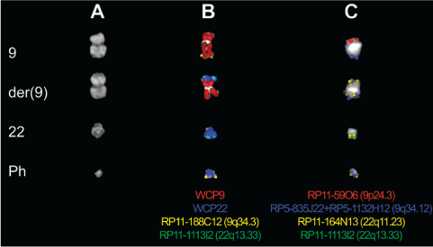
    Figure 1:  Partial metaphases showing karyotype and FISH cohybridization
experiments performed on the case under study. Each column shows normal
and derivative chromosomes 9 and 22. Whole Chromosome Paints (WCP),
Bacterial Artificial Chromosome (BAC) and P1-derived Artificial Chromosome
(PAC) probes shown at the bottom of each column are indicated with the
same pseudocolor in the merged images. A) Partial Q-banded karyotype. B)
Co-hybridization FISH experiments using the WCP probes of chromosomes
9 (red) and 22 (blue), as well as BAC clones mapping at the subtelomeric
region of the long arm of chromosomes 9 (yellow) and 22 (green). C) FISH
experiments performed to map the breakpoints on both the short [RP11-59O6
(red)] and long [RP11-164N13 (yellow)] arm of the inverted der(9). The two
PAC clones used as a pool probe, encompassing the ABL1 gene, are shown
in blue.
FISH experiments were performed as previously described [5] by co-hybridizing the Whole Chromosome Painting (WCP) probes as well as the subtelomeric Bacterial Artificial Chromosome (BAC) (Roswell Park Cancer Institute [RPCI]-11 Human Male Bac Library, Buffalo, NY) of the long arm of chromosomes 9 and 22 (RP11- 188C12 and RP11-1113I2, respectively), together with the P1-derived Artificial Chromosome (PAC) (Roswell Park Cancer Institute [RPCI]-5 Human Male PAC Library, Buffalo, NY) encompassing the ABL1 gene. The results revealed that the additional material on der(9) derived from chromosome 22 (Figure 1B). We hypothesized the occurrence of a paracentric inversion, leading to the repositioning of the chromosome 22 translocated material from the long to the short arm of der(9). Subsequent FISH investigations, using appropriate BAC clones, mapped the breakpoint on the short arm of der(9) within the overlapping region between RP11-59O6 and RP11-620I15 clones, encompassing the coding sequence of the Dedicator of Cytokinesis 8 (DOCK8) gene. On the translocated chromosome 22, the breakpoint was located within the BAC clone RP11-164N13 containing the BCR gene (Figure 1C). Of note, this clone showed, apart from the expected splitting signals on the Ph chromosome and the long arm of der(9), also an extra signal on the short arm of der(9) (Figure 1C). Thus, the rearrangement, not accompanied by any copy number alteration (as observed by SNP array analysis; Genome-Wide Human SNP Array 6.0, Affymetrix; Supplementary Table 1), led to the juxtaposition of DOCK8 and BCR with an opposite transcriptional orientation, without the genesis of any fusion transcript.
To assess the possible impact of this rearrangement upon DOCK8, Reverse Transcriptase quantitative PCR (RT-qPCR) experiments were performed, as previously described [6], on the patient’s Bone Marrow (BM) RNA. The mean gene expression value in three CML Ph+ control cases was used as calibrator and the geometric mean of GAPDH, ACTß e 28S genes as reference. Commercially available RNA (Clontech, Jesi, Italy) from BM (Cat. No. 636591) was used as normal control. According to the GRCh37/hg19 sequence assembly of the UCSC Human Genome Browser, DOCK8 displayed ten different isoforms, whose the longest one contained 48 exons (Figure 2A). A total of nineteen exons were tested by properly designed primer pairs along the inverted (two primer pairs: EX I-II and EX IV-V), the breakpoint (two primer pairs: EX VI-VII and EX XIII-XIV), and the retained portion of the gene (six primer pairs: EX XX-XXI, EX XXVI, EX XXXI-XXXII, EX XXXVI-XXXVII, EX XLI-XLII, and EX XLVXLVI) (all primer sequences are available upon request) and used in RT-qPCR experiments. Statistical data analysis, performed using the Relative Expression Software Tool (REST) [7], showed that there was no significant change in the expression of DOCK8, at least for the exons investigated, in the patient under study when compared with the mean Ct value of controls (Figure 2B).
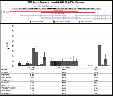
    Figure 2:  Expression analysis of DOCK8. A) UCSC Human Genome Browser
(GRCh37/hg19) map of the DOCK8 gene in chromosome band 9p24.3. The
clones in red showed a splitting signal on both the short and long arm of der
(9) in FISH experiments, and thus identified the breakpoint region. B) RTqPCR
results of the tested nineteen exons of DOCK8, mapping within the
inverted (bars with black vertical lines), breakpoint (bars with black horizontal
lines), and retained region (bars in grey), of the gene in the present case
(CML case), in 3 CML control cases (CML mean value), and in normal Bone
Marrow (BM).
Discussion/Conclusion
In summary, we here described for the first time a novel paracentric inversion affecting DOCK8 on the der(9) chromosome in a Ph+ CML patient, without originating any fusion transcript.
DOCK8 encodes for a member of the DOCK180 super family of guanine nucleotide exchange factors, which interacts with Rho GTPases. The protein is a component of intracellular signaling networks that regulate actin cytoskeletal rearrangement, cell migration, cell morphology, growth, proliferation, and differentiation [8]. DOCK8 inactivation due to deletion or mutation was previously linked to both mental retardation and autism [9], as well as to combined immunodeficiency syndromes [10]. According to the literature, patients with immunodeficiency syndromes and DOCK8 deficiency showed an increased susceptibility to specific infections and cancer development [8-10]. This is probably caused by a defective activation and differentiation of CD8+ T-cells, which are crucial component of the cellular immune response [11]. Interestingly, homozygous DOCK8 deletions were found in many human tumors, such as lung cancer [12], low-grade gliomas [13], hepatocellular carcinoma [14], neuroblastoma [15], cutaneous T-cell lymphoma/ leukemia [10], and Burkitt lymphoma, suggesting a possible role of this gene as a tumor suppressor.
In the present case, since the driver mutation leading to leukemia development is the BCR-ABL1 fusion gene, we speculate that the rearrangement involving DOCK8 should be a secondary event following the canonical t(9;22) translocation. In our hands, it was not possible to clearly define the clinical impact of this genomic alteration, due to the absence of intra-genic deletions as well as to the lack of this gene dysregulation (at least for 19 out of the 48 exons tested by RT-qPCR). However, we could not exclude that the fusion of BCR sequence to DOCK8 might have produced alternative DOCK8 splicing variants encoding for truncated protein isoforms. This variants might contribute to leukemogenesis acting as dominant negative inhibitors of the wild-type protein, similarly to what reported for RUNX1 [16] and PAX5 [17] tumor suppressor genes. Therefore, the study of further CML cases with DOCK8 rearrangements would allow a better understanding of the clinical and molecular impact upon leukemogenesis of this gene.
References
- Chereda B, Melo JV. Natural course and biology of CML. Ann Hematol. 2015; 94: S107-121.
- Jabbour E, Kantarjian H. Chronic myeloid leukemia: 2014 update on diagnosis, monitoring, and management. Am J Hematol. 2014; 89: 547-556.
- Vardiman JW. The World Health Organization (WHO) classification of tumors of the hematopoietic and lymphoid tissues: an overview with emphasis on the myeloid neoplasms. Chem Biol Interact. 2010; 184: 16-20.
- Erba HP. Molecular monitoring to improve outcomes in patients with chronic myeloid leukemia in chronic phase: importance of achieving treatment-free remission. Am J Hematol. 2015; 90: 242-249.
- Storlazzi CT, Albano F, Dencic-Fekete M, Djordjevic V, Rocchi M. Late-appearing pseudocentric fission event during chronic myeloid leukemia progression. Cancer Genet Cytogenet. 2007; 174: 61-67.
- Storlazzi CT, Fioretos T, Surace C, Lonoce A, Mastrorilli A, Strombeck B, et al. MYC-containing double minutes in hematologic malignancies: evidence in favor of the episome model and exclusion of MYC as the target gene. Human molecular genetics. 2006; 15: 933-942.
- Pfaffl MW, Horgan GW, Dempfle L. Relative expression software tool (REST) for group-wise comparison and statistical analysis of relative expression results in real-time PCR. Nucleic Acids Res. 2002; 30: e36.
- Rezaei N, Hedayat M, Aghamohammadi A, Nichols KE. Primary immunodeficiency diseases associated with increased susceptibility to viral infections and malignancies. The Journal of allergy and clinical immunology. 2011; 127: 1329-1341.
- Griggs BL, Ladd S, Saul RA, DuPont BR, Srivastava AK. Dedicator of cytokinesis 8 is disrupted in two patients with mental retardation and developmental disabilities. Genomics. 2008; 91: 195-202.
- Zhang Q, Davis JC, Lamborn IT, Freeman AF, Jing H, Favreau AJ, Matthews HF. Combined immunodeficiency associated with DOCK8 mutations. N Engl J Med. 2009; 361: 2046-2055.
- Lambe T, Crawford G, Johnson AL, Crockford TL, Bouriez-Jones T, Smyth AM, Pham TH. DOCK8 is essential for T-cell survival and the maintenance of CD8+ T-cell memory. Eur J Immunol. 2011; 41: 3423-3435.
- Takahashi K, Kohno T, Ajima R, Sasaki H, Minna JD, Fujiwara T, et al. Homozygous deletion and reduced expression of the DOCK8 gene in human lung cancer. Int J Oncol. 2006; 28: 321-328.
- Idbaih A, Carvalho Silva R, Crinière E, Marie Y, Carpentier C, Boisselier B, et al. Genomic changes in progression of low-grade gliomas. J Neurooncol. 2008; 90: 133-140.
- Saelee P, Wongkham S, Puapairoj A, Khuntikeo N, Petmitr S, Chariyalertsak S, et al. Novel PNLIPRP3 and DOCK8 gene expression and prognostic implications of DNA loss on chromosome 10q25.3 in hepatocellular carcinoma. Asian Pacific journal of cancer prevention: APJCP. 2009; 10: 501-506.
- Schramm A, Köster J, Assenov Y, Althoff K, Peifer M, Mahlow E, et al. Mutational dynamics between primary and relapse neuroblastomas. Nat Genet. 2015; 47: 872-877.
- Rodriguez-Perales S, Torres-Ruiz R, Suela J, Acquadro F, Martin MC, Yebra E, et al. Truncated RUNX1 protein generated by a novel t(1;21)(p32;q22) chromosomal translocation impairs the proliferation and differentiation of human hematopoietic progenitors. Oncogene. 2015; 70.
- Coyaud E, Struski S, Prade N, Familiades J, Eichner R, Quelen C, et al. Wide diversity of PAX5 alterations in B-ALL: a Groupe Francophone de Cytogenetique Hematologique study. Blood. 2010; 115: 3089-3097.