
Case Report
J Blood Disord. 2017; 4(1): 1044.
Microangiopathic Hemolytic Anemia with Stomach Cancer
Singla V1, Kunal D2*, Kala M3 and Verma SK4
1Department of Radiation Oncology, Swami Rama Himalayan University, India
2Division of Hematology & Pediatric Oncology, Swami Rama Himalayan University, India
3Department of Pathology, Swami Rama Himalayan University, India
4Division of Medical Oncology, Swami Rama Himalayan University, India
*Corresponding author: Kunal D, Division of Hematology & Pediatric Oncology, Swami Rama Himalayan University, India
Received: March 17, 2017; Accepted: April 11, 2017; Published: April 18, 2017
Abstract
Microangiopathic Hemolytic Anemia [MAHA] is a rare hematological condition due to red blood cell breakdown. Various etiologies has been noted, however its occurrence in oncological condition is sparse. Carcinoma stomach associated with MAHA is generally having fulminant course. Detection of MAHA at diagnosis of malignancy has been noted earlier as well. We encountered a case of carcinoma stomach that developed MAHA during early treatment phase and had short survival.
Keywords: Microangiopathic hemolytic anemia; Carcinoma stomach; Signet cell; Cancer-related hemolytic anemia
Introduction
Microangiopathic Hemolytic Anemia (MAHA) has been characterized by hemolysis due to non-immune lysis of RBC. Presence of dysmorphic fragmented red blood cells and schistocytes on peripheral smear are indicative of this entity. Associated thrombocytopenia is also noted. It has been associated with variable physiological (pregnancy induced) or pathological conditions (thrombotic thrombocytopenic purpura, hemolytic uremic syndrome, malignancy associated, hematological diseases) [1]. Among malignant conditions, association with carcinoma stomach, lung, breast, unknown primary and lymphoma are common [2]. We describe a case report of patient with carcinoma stomach who developed MAHA during the course of treatment and had rapid downhill course.
Case Summary
A 45 year old man was admitted with recurrent vomiting and abdominal discomfort for 4 weeks. Physical examination showed an ECOG performance status of III. Systemic examination revealed mild abdominal distension with a vague lump in upper half of abdomen. There was no palpable peripheral lymphadenopathy or hepato-splenomegaly. He was evaluated with endoscopy and found to have ulcerative growth in distal stomach. Biopsy was suggestive of carcinoma stomach with signet rings. Immunohistochemistry was positive for cytokeratin and negative for CK7, CK20, Napsin A, TTF, Synaptophysin, Hep-par 1, p63, PLAP and AFP confirming the diagnosis of adenocarcinoma stomach. PET CT evaluation showed stage IV disease involving thoracic and abdominal nodes as well as diffuse involvement of bone marrow. Bone marrow biopsy confirmed diffuse disease infiltration. He was started on chemotherapy [capecetabine-oxaloplatin]. He became asymptomatic after first cycle but developed acute onset progressive anemia and jaundice before second due course. Evaluation showed indirect hyperbilirubinemia with anemia and thrombocytopenia. Peripheral smear showed marked aniso-poikilocytosis with schistocytes (Figure 1). Direct coombs test was negative. Serum creatinine was normal and urine analysis showed hemoglobinuria. He was diagnosed to have microangiopathic hemolytic anemia with thrombocytopenia and started on steroid [methylprednisolone 1gm IV x 3 days]. Transfusions of packed red cell and platelets were done but showed no improvement in blood parameters. Intravenous immunoglobulin [IVIG 1gm/kg infusion] was administered along with fresh frozen plasma which showed transient improvement in blood parameters. His chemotherapy was held as Oxaliplatin induced MAHA was considered differential. However hemolysis along with thrombocytopenia continued and patient developed one episode of brief unconsciousness. MRI brain showed nodular lesion in left parietal cortex, possibly disease metastasis. A rapidly progressing disease with cancer related MAHA was considered and he was restarted on chemotherapy with steroid and IVIG dosing. However his hematological parameters remained deranged with persistent hemolytic anemia and thrombocytopenia. He became symptomatic for increased abdominal mass as well. He was transfused with packed red cells and platelet pharesis units under steroid cover but showed no response. He developed fever and decreased cognition. Family opted for supportive terminal care and he succumbed to his ailments in next 3 days.
Discussion
Metastatic signet ring cell carcinoma has been sparsely associated with MAHA. Reports of MAHA as first clinical feature leading to diagnosis of underlying malignancy are noted in literature [3]. Hahn et al reported a very high incidence (25.5%) of underlying carcinoma stomach in MAHA cases; however similar incidences were not noted elsewhere [4]. However a thorough search of malignancy, including bone marrow examination should be done in an unexplained case of MAHA. Index case developed MAHA during early phase of treatment. Absence of MAHA during diagnosis and first chemotherapy was unusual. A chemotherapy induced MAHA was considered as differential however further course with rapid clinical worsening was more suggestive of cancer related MAHA [CR-MAHA].
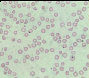
Figure 1: Peripheral smear showing marked aniso-poikilocytosis with schistocytes.
Exact pathogenesis of CR-MAHA is not known, however cancer related bone marrow necrosis and micro embolism has been postulated. Pro-coagulants and tumor related factors as well as lysis by-products have been implicated in causation [5]. A decrease in level of ADAMTS13 has also been demonstrated in CR-MAHA cases [6]. Index case had diffuse bone marrow involvement but no significant necrosis of marrow was noticed. CR-MAHA has been shown response to the treatment of primary malignancy. For usual MAHA, steroid, Rituximab, immunoglobulin and plasmapheresis have been noted to be beneficial [7]. Index case was treated with steroid and later on Immunoglobulin was administered as well. Fear of Rituximab induced immune suppression in post chemotherapy setting precluded its use. He showed short lasting partial response only.
Cancer related MAHA [CR-MAHA] is usually associated with aggressive course of disease. For even stage IV malignancy, occurrence of MAHA has been found to be predictor of worse prognosis in comparison to no MAHA cases [8]. Most of CR-MAHA cases die within few weeks, majority with infection as final insult [3,4,6]. Index case also developed rapid downhill course post CR-MAHA and died within 6 weeks of diagnosis.
CR-MAHA is an uncommon para-neoplastic syndrome with rapidly fatal outcome. Its association with carcinoma stomach and specially signet ring cell variant confer a worse prognosis. Aggressive tumor control is possibly the most effective treatment for CR-MAHA as well.
References
- George JN, Charania RS. Evaluation of patients with microangiopathic hemolytic anemia and thrombocytopenia. Semin Thromb Hemost. 2013: 39: 153-160.
- Antman KH, Skarin AT, Mayer RJ, Hargreaves HK, Canellos GP. Microangiopathic hemolytic anemia and cancer: A review. Medicine. 1979; 58: 377-384.
- Shin SY, Park H, Chae SW, Woo HY. Microangiopathic hemolytic anemia as the first manifestation of metastatic signet ring cell carcinoma of unknown origin: a case report and review of literature. Korean j Lab Med. 2011; 31: 157-161.
- Hahn JS, Lee DH, Lee SJ, Min YH, Ko YW. A clinical study on microangiopathic hemolytic anemia. Korean J Hematol. 1991; 26: 263-279.
- Caine GJ, Stonelake PS, Lip GY, Kehoe ST. The hypercoagulable state of malignancy: pathogenesis and current debate. Neoplasia. 2002; 4: 465-473.
- Francis KK, Kalyanam N, Terrell DR, Vesely SK, George JN. Disseminated malignancy misdiagnosed as thrombotic thrombocytopenic purpura: A report of 10 patients and a systemic review of published cases. Oncologist. 2007; 12: 11-19.
- Espingosa G, Bucciarelli S, Cervera R, Lozano M, Reverter JC, de la Red G, et al. Thrombotic microangiopathic hemolytic anemia and antiphospholipid antibodies. Ann Rheum Dis. 2004; 63: 730-736.
- Etoh T, Baba H, Taketomi A, Nakashima H, Kohnoe S, Seo Y, et al. Diffuse bone metastasis with hematologic disorders from gastric cancer: clinicopathological features and prognosis. Oncol Rep. 1999; 6: 601-605.