Case Report
Austin J Cancer Clin Res 2015;2(4): 1037.
Endobronchial and Brain Metastases of Malignant Melanoma during an 11-Year Follow up: A Case Report and Review of the Literature
Yamauchi A1,2, Yokoyama Y2*, Morikawa A1, Soma T1, Ota K1, Yokota M1, Matsukura D1, Sato S1 and Mizunuma H2
1Department of Pulmonology, Instituto Nacional de Silicosis (INS), University of Oviedo, Spain
2Department of Pathology, Hospital Universitario Central de Asturias (HUCA), Spain
*Corresponding author: Ariza MA, Department of Pulmonology, Instituto Nacional de Silicosis (INS), Área del Pulmón, Hospital Universitario Central de Asturias (HUCA), University of Oviedo, Avenida Roma s/n, Oviedo, Asturias 33011, Spain.
Received: March 27, 2015; Accepted: May 07, 2015; Published: May 30, 2015
Abstract
Although lung parenchyma is a common site for metastasis from extrathoracic tumors including melanoma, endobronchial metastasis from malignant melanoma is a very rare phenomenon. Malignant melanoma is generally known as a rapidly growing tumor, and recurrences are often observed within a short period. The most frequent non-pulmonary primary tumors with endobronchial metastasis are breast, kidney and colon. Endobronchial metastasis from malignant melanoma can simulate bronchogenic carcinoma and presenting symptoms include cough, hemoptysis, dyspnea and wheezing. There are only a few reports of endobronchial metastasis from malignant melanoma. Compared with primary lung, breast, renal or colorectal cancer, melanoma has the highest propensity to metastasize to the brain and these patients have significantly the worst overall prognosis. We report the first known described case of a 42-yearold woman who was diagnosed with endobronchial and brain metastases from a left thigh skin melanoma after being disease-free for 11 years.
Keywords: Malignant melanoma; Endobronchial; Pulmonary; Brain; Metastasis
Abbreviations
CT: Computed Tomography; LDH: Lactic Dehydrogenase; MRI: Magnetic Resonance Imaging; MM: Malignant Melanoma; CECT: Contrast-enhanced Computed Tomography; PET: Positron Emission Tomography
Case Presentation
A 42-year-old woman had complained of a black skin lesion on the inner face and proximal third of her left thigh, which was diagnosed as nodular melanoma in February 1997. She underwent a left inguinal linfadenectomy and excision of the tumor with 3-cm margins with no evidence of residual melanoma. The patient remained on routine follow-up visits until 2007. She had a 20 pack-year history of smoking and no other surgical or other medical background of interest. After 11 years of disease-free, the patient was admitted in our hospital, in February 2008, after a week with dizziness and walking instability accompanied by occipital headache, persistent nausea and bilious vomit. She also referred non productive cough in the last month with no chest pain, night sweats or fever. During the neurological examination, a vertical nystagmus on upgaze and a finger-nose-finger dysmetria were seen. The pulmonary auscultation was normal. She was taken only antiemetic drugs at the time.
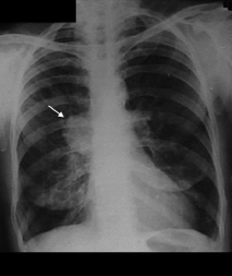
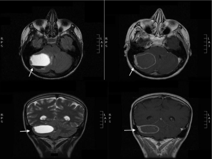
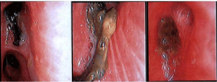
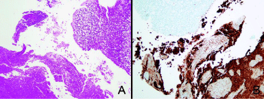
A cranial computed tomography (CT) scan was performed. The cranial CT revealed a low intensity mass in the right cerebellar hemisphere with an important mass effect, which compressed the fourth ventricle and showed contrast uptake in the form of ¨ring enhancement¨. The chest X-ray showed a right parahilar mass with signs of postobstructive pneumonitis (Figure 1). Laboratory tests (complete blood count, biochemical and arterial blood clotting) were within normal ranges, except for lactic dehydrogenase LDH of 802 IU/L. Three repeated sputum cytologic examinations were negative for malignant cells. The magnetic resonance imaging (MRI) of the brain showed a right cerebellar hemisphere hemorrhagic lesion with an important associated mass effect with lower displacement of the cerebellar tonsils and partial collapse of the fourth ventricle with perilesional enhancement of the mass after contrast administration (Figure 2). Bronchoscopy showed a black-colored endobronchial mass in the right upper lobe causing complete obstruction of the anterior segment (Figure 3). The pathological examination of the bronchoscopy biopsy revealed the presence of malignant melanoma cells. Immunohistochemical analysis revealed the tumor cells were positive for both HMB45 (gp100) and MART-1 (Melan A) (Figure 4). The final diagnosis was endobronchial metastasis of malignant melanoma. In the sixth day of admission, the patient was in a very delicate clinical situation with an important neurological deterioration. She was examined by the neurosurgery department, but due to the poor short term outcome and in accordance with her family, aggressive treatment was discarded. Unfortunately the patient died on the sixth day of admission.

    Figure 1:  A) A small fragment of benign respiratory epithelium (upper right)
surrounded by necrotic tissue, H/EX200. (B) Immunohistochemistry revealed
that much of the necrotic cells were positive for HMB45 antibody (melanoma
cells), peroxidase X 200.

    Figure 2:  Brain magnetic resonance imaging (MRI): Right cerebellar
hemisphere hemorrhagic lesion with lower displacement of the cerebellar
tonsils and partial collapse of the fourth ventricle with perilesional
enhancement of the mass after contrast administration.

    Figure 3:  Fiberoptic bronchoscopy showing a darkly pigmented lesion of
endobronchial melanoma in the right upper lobe causing complete obstruction
of the previous segment.

    Figure 4:  A) A small fragment of benign respiratory epithelium (upper right)
surrounded by necrotic tissue, H/EX200. (B) Immunohistochemistry revealed
that much of the necrotic cells were positive for HMB45 antibody (melanoma
cells), peroxidase X 200.
Discussion
Malignant melanoma (MM) is a tumor that arises from the pigment producing cells (melanosomes) of the deeper layers of the skin (or the eye) and is the leading cause of death attributable to skin lesions. It is usually described as an irregular dark skin lesion that may have areas of varying colour. Although malignant melanoma metastasizes to several organs, endobronchial metastasis is rare [1]. Furthermore, MM is generally known as a rapidly growing tumor, and recurrence are often observed within a short period [2]. Thus, it is quite unusual for metastasis to be observed during a prolonged clinical course as seen in our case. Endobronchial metastases from non-pulmonary primary malignancies are rare, occurring in less than 2% of patients [3]. The most frequent non-pulmonary primary tumors with endobronchial metastasis are breast, kidney and colon [4]. When cutaneous melanoma disseminates, it has no preferential pattern of metastasis [5]. In a review from 1962-2002, Sorensen, found a total of 204 patients with endobronchial metastases originating from 20 different primary extrapulmonary tumors [6]. In this review, 8 of the cases were caused by metastatic melanoma.
We used PubMed to search for literature on endobronchial metastasis from skin melanoma with the following index words: endobronchial melanoma metastasis, endobronchial and brain melanoma metastasis. A total of only 15 clinical cases were found in the search, we did not find any case of both endobronchial and brain melanoma metastases in the same patient as seen in our report. Endobronchial metastasis most commonly present with cough, shortness of breath, and hemoptysis [6]. Our patient presented with dry cough (the only respiratory symptom). The bronchoscopy in our patient showed a right upper lobe mass with black pigmentation obstructing completely the anterior segment. Black pigmentation in the airways is a benign finding in most patients, but it can be the sign of systemic disease or rare malignancy in some patients. There are many causes of black pigmentation in the airways: anthracosis, systemic diseases like alkaptonuria, argyrosis, iron overload, amiodarone toxicity, and charcoal aspiration have all been reported as unusual causes of black airway pigmentation [7]. Melanoma can have the appearance of a black infiltrating endobronchial mass or plaque in the airways [8]. The term ¨black bronchoscopy¨ was first coined by Packham et al. [9] to describe a case of endobronchial metastasis in malignant melanoma from an ear. In our patient, a history of thigh skin melanoma made us suspect the diagnosis of endobronchial metastasis. However, histologic finding of malignant cells containing melanin pigment is critical to confirm the diagnosis, given that there are a number of possible differential diagnoses for the presence of black pigmentation in the airways.
Our patient symptoms were mainly neurological, she only referred cough as the only respiratory symptom. As evident in our case, the time of appearance of endobronchial metastasis can be long after the diagnosis of primary tumor. A carefully, taken history is crucial as it helps in the initial approach to the patient´s symptoms. It also aids in differentiating endobronchial metastasis from primary bronchogenic tumors, which can have a similar clinical presentation. It is difficult to distinguish primary malignant melanoma of the lung from metastatic melanoma. The diagnostic criteria for primary malignant melanoma of the lung based on pathological findings include a functional change with a ¨dropping off¨ or ¨nesting¨ of malignant cells containing melanin just beneath the bronchial epithelium; invasion of the bronchial epithelium by melanoma cells in an area where the bronchial epithelium is damaged; and an obvious melanoma beneath an epithelium with these changes [10].
The high incidence of brain metastasis in melanoma leads to a high frequency of inclusion of brain lesions in staging, and it is not unusual to identify small asymptomatic lesions during staging. When symptoms occur, they are nonspecific and vary depending on the location of the lesion. Headache is the most common presenting symptom, but patients may also present with more serious symptoms like seizures, hemiplegia or visual compromise due to raised intracranial pressure. These phenomena may suggest large (>4 cm) lesions, numerous lesions or frequently, tumoral hemorrhage which must be considered in patient management decisions [11].
Our patient referred headache, walking instability, and fingernose- finger dysmetria accompanied vertical nystagmus in the neurological examination. Patients with brain metastasis have significantly worse progression-free survival, overall survival and overall prognosis than patients with no brain metastasis. In our case, the patient started with headaches 2 weeks before admission, by that time, only palliative treatment could be offered. The mean survival after endobronchial metastasis is less than 6 month. CT findings of metastatic melanoma to the lungs include pulmonary nodules (occasionally in a miliary pattern), feeding vessels associated hematogenous lung metastases, postobstructive atelectasis, and hilar/ mediastinal lymphadenopathy [12]. Staging work-up of metastatic melanoma includes brain magnetic resonance imaging and either contrast-enhanced computed tomography (CECT) or positron emission tomography (PET)-CT of the chest, abdomen, and pelvis. PET-CT is more sensitive (86%) and specific (91%) than CECT (63% sensitivity and 78% specificity) in detecting melanoma metastases; however, due to the relatively reduced availability of PET imaging, CECT is often the study of choice [12].
Treatment for endobronchial metastasis has changed over the years as novel endobronchial therapies have emerged. In general, assessing the efficacy of various therapies is difficult because most subjects with endobronchial metastasis have diffuse metastatic disease and treatment is usually palliative. Older therapies included combined chemotherapy and radiation therapy with or without surgery. Newer modalities include the use of laser evaporation [13,14], endobronchial brachytherapy, stenting, and photodynamic therapy [15].
We believe metastatic melanoma should be included in the differential diagnosis for patients with endobronchial lesions, and clinicians should be aware of the risk of recurrence in patients with malignant melanoma, even in patients who remain in stable condition without recurrence for a prolonged period, as seen in our case.
References
- Akoglu S, Uçan ES, Celik G, Sener G, Sevinç C, Kilinç O, et al. Endobronchial metastases from extrathoracic malignancies. Clin Exp Metastasis. 2005; 22: 587-591.
- Sutton FD Jr, Vestal RE, Creagh CE. Varied presentations of metastatic pulmonary melanoma. Chest. 1974; 65: 415-419.
- Braman SS, Whitcomb ME. Endobronchial metastasis. Arch Intern Med. 1975; 135: 543-547.
- Meyer T, Merkel S, Goehl J, Hohenberger W. Surgical therapy for distant metastases of malignant melanoma. Cancer. 2000; 89: 1983-1991.
- Devita VT. Cutaneous melanoma. Cancer principles and practices of oncology 2nd Ed. 1985; 1371-1422.
- Sørensen JB. Endobronchial metastases from extrapulmonary solid tumors. Acta Oncol. 2004; 43: 73-79.
- Teo YK, Kor AC. "Black bronchoscopy"-a case of endobronchial metastases from melanoma. J Bronchology Interv Pulmonol. 2010; 17: 146-148.
- Das RK, Dasgupta A, Tewari S. Malignant melanoma of the bronchus. J Bronchol. 1998; 5: 59-60.
- Packham S, Jaiswal P, Kuo K, Goldsack N. Black bronchoscopy. Respiration. 2003; 70: 206.
- Allen MS Jr, Drash EC. Primary melanoma of the lung. Cancer. 1968; 21: 154-159.
- Goulart CR, Mattei TA, Ramina R. Cerebral melanoma metastases: a critical review on diagnostic methods and therapeutic options. ISRN Surg. 2011; 2011: 276908.
- Patnana M, Bronstein Y, Szklaruk J, Bedi DG, Hwu WJ, Gershenwald JE, et al. Multimethod imaging, staging, and spectrum of manifestations of metastatic melanoma. Clin Radiol. 2011; 66: 224-236.
- Capaccio P, Peri A, Fociani P, Ferri A, Ottaviani F. Flexible argon plasma coagulation treatment of obstructive tracheal metastatic melanoma. Am J Otolaryngol. 2002; 23: 253-255.
- Andrews AH Jr, Caldarelli DD. Carbon dioxide laser treatment of metastatic melanoma of the trachea and bronchi. Ann Otol Rhinol Laryngol. 1981; 90: 310-311.
- Strazel H. Fractionated intraluminal HDR 192lr brachytherapy as palliative treatment in patients with endobronchial metastases from non-bronchogenic primaries. Strahlenther Onkol, 2002; 178: 442-445.