
Clinical Image
Austin J Cancer Clin Res 2015;2(4): 1039.
Large Right Ventricular Metastasis of Melanoma
Banzato A¹*, Bianchi A¹, Pigozzo J² and Denas G¹
¹Cardiology Unit, Veneto Institute of Oncology IOV, IRCCS, Italy
²Melanoma and Esophageal Oncology Unit, Veneto Institute of Oncology IOV, IRCCS, Italy
*Corresponding author: Banzato A, Cardiology Unit, Veneto Institute of Oncology IOV, IRCCS, Via Gattamelata, 64, 35100 Padua, Italy.
Received: April 16, 2015; Accepted: June 14, 2015; Published: June 30, 2015
Clinical Image
A 62-year-old female was referred for congestive heart failure. Clinical history included diffuse malignant melanoma and no previous heart disease. She was in poor general conditions and lamented dyspnea, with no other cardiovascular symptoms. Physical examination showed jugular turgor, normal heart sounds without murmurs, lower limb edema and hepatomegalia with pronounced hepatojugular reflux. The ECG showed sinus rhythm, Q waves in the inferior leads and diffuses T-wave inversion (panel A). Differential diagnosis included: pulmonary embolism, cardiotoxicity from chemotherapy, pericardial effusion but even unrecognized myocardial infarction. Echocardiogram is the cornerstone for differential diagnosis. What we found was none of our suppositions, and it is depicted in panel B: a large mass occupying almost the whole right ventricle growing from the apex to just above the tricuspid valve. Because there was no obstruction in right ventricular inflow and outflow, the condition was not lethal. New cancer treatment has improved patient longevity, so is more frequent to meet cardiac metastasis.
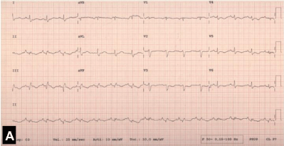
    Figure 1:  A large mass at whole right ventricle growing from the apex to just
above the tricuspid valve.