Clinical Image
Austin J Cancer Clin Res 2015;2(6): 1047.
Pleurx Exit Site Metastasis in Squamous Cell Lung Cancer
Friend R* and Walker P
Department of Hematology & Oncology, East Carolina University, USA
*Corresponding author: Reed Friend, Department of Hematology & Oncology, East Carolina University, Grenville, NC, USA.
Received: May 05, 2015; Accepted: June 06, 2015; Published: July 28, 2015
Clinical Image
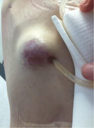
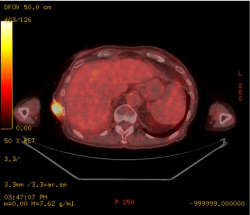
A 69-year-old female, who wasone month post definitive chemoradiation for a Stage IIIB Squamous cell carcinoma of the lung, reporteda new nodule at her Pleurx catheter exit site (Figure 1). This lesion was confirmed on fine needle aspiration to be metastatic squamous cell lung cancer consistent with the primary diagnosis. Pleural fluid cytology was negative for malinant cells on two separate analyses, one month apart. Positron emission tomography (PET) restaging showed marked FDG avidity at the Pleurx exit site nodule (Figure 2) without evidence of a pleural effusion or pleural thickening. Seeding along the tract of a pleurx catheter has been reported in cases where there is a confirmed malignant pleural effusion and evidence of seeding [1]. This is, to our knowledge, the only case reported of a spontaneous subcutaneous metastatic lesion to arise at a catheter exit site without evidence of seeding.

Figure 1: Violacious non-tender nodule at Pleurx exit site.
