
Case Report
Austin Cardio & Cardiovasc Case Rep. 2017; 2(1): 1014.
Assessment of the Morphologic Right Ventricular Function after the Atrio-Ventricular Valve Replacement of a Corrected Transposition of the Great Arteries
Uchida W*, Mutsuga M, Hirate Y, Kawaguchi O and Usui A
Department of Cardiac Surgery, Nagoya University Graduate School of Medicine, Japan
*Corresponding author: Wataru Uchida, Department of Cardiac Surgery, Nagoya University Graduate School of Medicine 65, Tsurumai-Cho, Syowa-Ku, Nagoya, Aichi, Japan
Received: February 09, 2017; Accepted: February 27, 2017; Published: March 01, 2017
Abstract
Here we assessed the morphologic right ventricular dysfunction in an adult after a corrected transposition of the great arteries; this dysfunction was a result of morphologic tricuspid valve regurgitation. A 52-year-old man presented with dyspnoea on exertion. Echocardiography showed severe morphologic tricuspid valve regurgitation. A morphologic tricuspid valve replacement procedure was performed using a superior septal approach. We selected a less invasive procedure for the deteriorated right ventricle and assessed the prognosis of the morphologic right ventricle using a cardiopulmonary exercise testing system.
Learning Objective: It has been reported that atrio-ventricular replacement was common procedure for atrio-ventricular regurgitation of a corrected transposition, but it had not been reported the concrete assessment of the deteriorated systemic ventricular. Thus, we showed in paper that cardiopulmonary exercise might be effective for the postoperative prognosis of a corrected transposition of the great arteries.
Keywords: Corrected transposition; Morphologic tricuspid valve regurgitation; Superior-septal approach; Cardiopulmonary exercise testing system
Abbreviation
BNP: Brain Natriuretic Peptide; CCTGA: Congenitally Corrected Transposition of the Great Arterie; CPX; Cardiopulmonary Exercise Testing System; SAVV: Systemic Atrio Ventricular Valve; SV: Systemic Ventricular; TR: Tricuspid valve Regurgitation; VO2: Oxygen Uptake; VE/VCO2: Minute Ventilation/Carbon Dioxide Production; VSD: Ventricular Septal Defect
Introduction
Congenitally Corrected Transposition of the Great Arteries (CCTGA) is a rare cardiac anomaly with an incidence of less than 1% in patients with congenital heart disease. Most cases are associated with Ventricular Septal Defect (VSD) as the most common then surgical procedure is performed in childhood [1,2]. In adult cases, this surgical procedure is often required for progressive Systemic Atrio Ventricular Valve (SAVV) regurgitation; however, the morphologic right ventricle remains exposed to the systemic circulation postoperatively. Deteriorating Systemic Ventricular (SV) function has an adverse effect on long-term outcomes. We managed a case of SAVV surgery for CCTGA and assessed the SV dysfunction using a cardiopulmonary exercise testing system (CPX).
Case Presentation
A 52-year-old man was admitted to our hospital with dyspnoea on effort. He had been followed-up after a corrected transposition of the great arteries. Physical examination revealed a systolic murmur over the chest. A chest X-ray showed an increased cardiopulmonary ratio of more than 60%. Laboratory study results showed that the Brain Natriuretic Peptide (BNP) levels had increased to 485pg/mL. Echocardiography demonstrated an SV ejection fraction of 52.4% with severe SAVV regurgitation and an expanded left atrium. The CPX showed that the peak Oxygen Uptake (peak VO2) and minute Ventilation/Carbon Dioxide Production (VE/VCO2) slope were 12.4 mL/kg/min and 40.0, respectively. He had operated for SAVV regurgitation. During this surgery, we did not perform a right-sided left atriotomy, but utilized a trans-septal incision to approach the SAVV. The trans-septal incision provided a good operative view of the SAVV. We performed a 29-mm mechanical valve replacement (Carbomedics Standard Mitral Heart Valve, SORIN, Milano, Italy) for the SAVV and a left atrial appendage plication (the surgical time was 174 minutes and the aortic cross-clamp time was 51 min). Postoperative echocardiography showed an SV ejection fraction of 37.5%. The BNP level improved to 201, peak VO2 improved to 16.2 and VE/VCO2 slope changed to 34.2. His dyspnoea on effort had improved, and the patient continued his cardiac rehabilitation. One year later, the BNP level, peak VO2 and VE/VCO2 slope had further improved to 157, 17 and 28.7, respectively.
Discussion
CCTGA is a rare cardiac malformation characterized by a combination of discordant atrioventricular and ventriculo-arterial connections [1]. The left atrium communicates with the morphologic right ventricle, which leads to the aorta. Therefore, although blood flows in a normal direction, it passes through the right ventricular chamber; this means that the morphologic tricuspid valve and right ventricle are exposed to the systemic circulation.
The SV tends to respond difficultly to exercise in comparison to a normal left ventricle. The myocyte organization is different between the right and left ventricles [1]. Graham and colleagues reported that the incidence of morphologic right ventricular dysfunction is increasing [2]. By forty-five years old, 56% of patients got SV dysfunction associated with VSD, morphologic TR and so on [3]. Abnormalities of the morphologic tricuspid valve have reportedly been observed in up to nine-tenths of patients with CCTGA [1]. The corrected transposition has grave concerns for the morphologic right ventricular dysfunction in not only preoperative but also postoperative status. Consequently, we paid attention not to prolong the cardiac ischemic time by using the trans-septal approach. The septal leaflet of the morphologic tricuspid valve is usually displaced inferiorly towards the cardiac apex; this is called ‘Epstein’s malformation’ [1]. This might make it difficult to repair the SAVV. Therefore, we did not utilize a right-sided left atrium incision and approached the SAVV with a superior-septal incision; this made it easier to perform the SAVV replacement.
In this case, the preoperative BNP level was 406 and SV dysfunction was observed. It is difficult, however, to evaluate the SV prognosis using only the BNP level in CCTGA patients. On the other hand, the CPX allow for the analysis of gas exchange at rest, during exercise and during recovery; it also provides breath-by-breath measures of VO2, carbon dioxide output (VCO2) and Ventilation (VE). The peak VO2 is the mark of the cardiac output during exercise, and the VE/VCO2 slope is the mark of the ventilation efficacy for CO2 ejection from the body. Both reflect the prognosis index of chronic heart failure [4,5]. Better SV prognosis is expected with improved VO2 and VE/VCO2. Exercise along with appropriate medications may lead to improved physical conditioning, which may improve the peak VO2 and VE/ VCO2 slope (Figure 1).
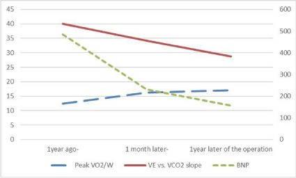
    Figure 1:  The VE/VCO2 slope directly correlated with the BNP level and
inversely correlated with the peak VO2/W. Association between Peak VO2,
VEVCO2 and BNP. Peak VO2, VE/VCO2 slopeand BNP were improved by
an operation and continued being improved 1 year later, respectively. Peak
VO2 was inversely correlated with VE/VCO2 slope and BNP.
We managed a case of an adult with CCTGA. To maintain the declining SV function, we believe that it is important to minimize damage through shortened surgical times and to evaluate the morphologic right ventricular prognosis using the CPX.
References
- Wallis GA, Debich-Spicer D, Anderson RH. Congenitally corrected transposition. Orphanet J Rare Dis. 2011; 6: 22.
- Graham TP Jr, Bernard YD, Mellen BG, Celermajer D, Baumgartner H, Cetta F, et al. Long-term outcome in congenitally corrected transposition of the great arteries: a multi-institutional study. J Am Coll Cardiol. 2000; 36: 255- 261.
- Bogers AJ, Head SJ, de Jong PL, Witsenburg M, Kappetein AP. Long term follow up after surgery in congenitally corrected transposition of the great arteries with a right ventricle in the systemic circulation. J Cardiothorac Surg. 2010; 5: 74.
- Ponikowski P, Francis DP, Piepoli MF, Davies LC, Chua TP, Davos CH, et al. Enhanced ventilatory response to exercise in patients with chronic heart failure and preserved exercise tolerance: maker of abnormal cardiorespiratory reflex control and predictor of poor prognosis. Circulation. 2001; 103: 967- 972.
- Opasich C, Pinna GD, Bobbio M, Sisti M, Demichelis B, Febo O, et al. Peak exercise oxygen consumption in chronic heart failure: toward efficient use in the individual patient. J Am Coll Cardiol. 1998; 31: 766-775.