
Review Article
Austin J Cardiovasc Dis Atherosclerosis. 2015; 2(1): 1012.
Women’s Heart Disease: A Review of the Current Testing Modalities for Coronary Microvascular Dysfunction
Pranav M. Patel MD* and Aaron Jolly MD
Division of Cardiology, Department of Medicine, University of California, Irvine, Orange, USA
*Corresponding author: Pranav M. Patel, Division of Cardiology, Department of Medicine, University of California, Irvine, Orange, USA
Received: March 17, 2015; Accepted: September 10, 2015; Published: September 25, 2015
Abstract
Up to 40% of female patients with chest pain syndromes are found to have non-obstructive coronary artery disease. Of these patients, studies have shown, an increase risk of poor cardiovascular outcomes, adverse prognosis and increased mortality. Coronary microvascular dysfunction has been proposed as the etiology to explain such symptoms and increased risks, especially in the female population. Several non-invasive and invasive techniques of assessing coronary microvascular dysfunction have been studied and have been shown to be safe. The use of coronary microvascular dysfunction testing in the cardiac catheterization laboratory by measuring coronary flow reserve (CFR) has been proven to be both sensitive and specific measure for microvascular disease, although it is not used on routine basis. This review of the literature describes the various microvascular disease testing available in current practice (especially in the female population). Such testing includes various protocols used in the cardiac catheterization lab to measure CFR.CFR, defined as the ratio of maximal coronary flow with hyperemia to baseline flow can be a routine testing modality in the cardiac catheterization laboratory. These types of protocols play an important component in the diagnosis of coronary microvascular dysfunction, and can be a routine testing protocol in cath labs that are willing to embrace such testing modalities.
Keywords: Heart disease; Microvascular disease; CFR; Microvascular dysfunction
Introduction
Coronary physiology dictates that coronary arterial vessels constantly adapt through vasomotor tone in response to changes in myocardial oxygen demand [1]. Lack of this ability to adapt to increasing myocardial oxygen requirements results in a mismatch between myocardial oxygen supply and demand, and can sometimes lead to myocardial ischemia. To prevent such ischemia the coronary arterial tree has blood flow reserve. This coronary blood flow reserve is defined as increase in blood flow in response to metabolic or pharmacological stimulations. Normal coronary blood flow reserve increases 2.5-5 fold in response to increases in oxygen demands [1].
In order to evaluate chest pain syndromes many patients undergo coronary angiogram in order to investigate for coronary artery disease. Patel et al, found that 39.2% of a study population of almost 400,000 patients, to have normal angiograms defined by <20% stenosis in coronary artery vessels. This study population was comprised of 47.3 % females. The percentage of patients who were female with obstructive coronary artery disease and non-obstructive coronary disease were 33.9% and 55.4% respectively [2]. Of these patients, studies have shown, women with impaired glucose tolerance or diabetes presenting with angina, regardless of angiographic findings, were found to have increased mortality [3]. The National Heart, Lung, and Blood Institute sponsored WISE (Women’s Ischemic Syndrome Evaluation) has shown that of symptomatic women (with normal angiograms after abnormal cardiac stress testing), up to one half have no obstructive coronary artery disease [4]. As a follow up to the WISE study population, Johnson et al found women with persistent chest pain with non-obstructive coronary artery disease predicted adverse cardiovascular outcomes [5]. Therefore, suggesting that undiagnosed disease likely contributes to anginal symptoms, adverse outcomes and mortality despite non-obstructive coronary artery disease by angiogram. Review of different methods to evaluate coronary flow reserve, concluded a reduction in coronary blood flow reserve in absence of obstructive flow-limiting coronary stenosis suggests vascular dysfunction as etiology of chest pain syndromes [6].
In 1985, Cannon and Epstein introduced a concept of “microvascular angina”. They proposed small vessel dysfunction might play a role in pathogenesis leading to chest pain syndromes [7]. Further studies have shown both coronary artery spasm and endothelial dysfunction to be predictors of morbidity and mortality in patients with angina [8, 9],
The combination of these findings led to several studies to evaluate the possibility of microvascular disease in symptomatic women with normal coronary arteries disease. In addition to microvascular disease as a diagnosis for angina in women with normal coronary anatomy, microvascular disease has been shown to be possibly present in patients diagnosed with takotsubo cardiomyopathy [10]. The distinction of cause-and-effect relationship has not be proven, alternative theories of microcirculation abnormalities as a result of wall stress from apical ballooning have been suggested [11].
Microvascular Disease Testing
Over a 20-year period several invasive and non-invasive techniques have been used for assessment of coronary microvascular dysfunction specifically with respect to impaired coronary blood flow reserve.
Coronary blood flow is regulated by different factors influencing both macrovascular and microvascular vasomotor tone. These include endothelium-dependent and independent factors [1]. The endothelium regulates vasomotor tone by stimulating release of vasoactive factors, specifically, nitric oxide, originally identified as endothelium-derived relaxing factor [12]. Various techniques have been used to replicate or simulate vasomotor tone in an effort to evaluate for microvascular dysfunction as a function of reduction of coronary flow reserve.
Useful non-invasive methods for diagnosis microvascular dysfunction as a reduction in coronary flow reserve include using positron emission tomography [13], cardiac magnetic resonance [14] and transthoracic Doppler echocardiography [15]. Invasive coronary vasomotor testing as shown by Wei et al, involves macrovascular and microvascular evaluation with both endothelium dependent and independent tests involving intracoronary injections of adenosine, acetylcholine and nitroglycerine [16]. Currently, invasive coronary vasomotor testing with acetylcholine remains the gold standard for diagnosis of coronary microvascular dysfunction with a welldescribed risk and benefit record [9, 16]. Furthermore, patients with a diagnosis of microvascular dysfunction are at increased risk for adverse outcomes over time [16].
Coffman and Gregg, in 1960, first introduced the idea of coronary arterial reactive hyperemia, which serves as the basis for many of these tests [17]. It has been established that vasodilator responses to acetylcholine occurs in normal coronary arteries through the release of endothelium-derived relaxing factors including nitric oxide [18]. Paradoxically the response was reversed to constriction of vessels in patients with evidence of coronary artery disease [19]. Intracoronary acetylcholine is considered the gold standard for detection of coronary endothelial function [20]. The safety of using intracoronary acetylcholine for the purpose of diagnosing microvascular disease has shown to have few adverse effects [9]. Most of these reactions were related to the effects of acetylcholine’s paradoxical constriction and spasm of the coronary arteries. Sueda et al showed 1.7% major adverse reaction out of 1000 patients tested, these ranged from non-sustained ventricular tachycardia, ventricular fibrillation, shock and cardiac tamponade [21]. Ong et al, in 2008, showed 86 patients with acute coronary syndrome, who underwent intracoronary acetylcholine infusion, had no adverse events documented [9]. As a result, many studies follow doses of acetylcholine with nitroglycerine to ensure dilatation of these vessels post vasoreactivity testing. The clinical measures for diagnosis of microvascular dysfunction following intracoronary acetylcholine have included coronary diameter reduction as well as coronary blood flow measurements derived from Doppler peak velocities.
Doppler of peak velocity with regards to coronary blood flow was evaluated post acetylcholine infusion using flow and pressure measurements using Doppler flow wires. Coronary blood flow changes < 50% at highest dose of acetylcholine was defined as an abnormal test result suggesting endothelial-dependent microvascular disease. This was followed by measurements of coronary artery dilation as a function of Doppler-derived velocity defined by < 5% changes as abnormal suggesting endothelial-dependent macrovascular disease. Coronary flow reserve (CRF) is defined as the ratio of maximal coronary flow with hyperemia to baseline flow. CFR tests the ability of epicardial stenosis and microcirculatory resistance to maintain flow. In the WISE study, 159 women with chest pain and non-obstructive coronary artery disease a coronary reserve velocity of 2.24 provided a 90% sensitivity and 89% specificity for the diagnosis of microvascular dysfunction based on a CFR of < 2.516.
Discussion
Evaluation of patient with chest pain syndromes typically involves possible stress test evaluation with definitive diagnosis of coronary artery disease by coronary angiogram to evaluate for obstructive macrovascular disease. Up to 40% of these patients have normal angiograms however this same subset has been shown to have higher risk for adverse cardiac events despite a normal angiogram [2-4]. These patients are told they have normal coronary arteries and advised further evaluation for non-cardiac etiologies for their symptoms.
Invasive testing to measure microvascular dysfunction includes CFR testing in the cath lab using intracoronary acetylcholine, adenosine and nitroglycerine. Coronary arterial blood flow velocity and coronary flow reserveare assessed using a 0.014-inch Doppler coronary wire with a piezoelectric ultrasound transducer at the wire tip to determine volumetric flow. The position of the Doppler sensor is in the proximal to mid portion of the vessel and manipulated until an optimal and stable blood flow velocity signal is obtained. The sensors are positioned at the same location for all measurements obtained throughout the procedure. Coronary flow reserve relates to the ability, in response to oxygen demands, of coronary vessels to increase blood flow to supply myocardium [22]. It is a ratio of maximal coronary blood flow to resting coronary blood flow. Bradley et al described the use of intracoronary Doppler flow meters to measure coronary blood flow velocity, which is proportional to flow [23]. Coronary flow reserve can be calculated as the ratio of resting compared to maximal blood flow velocity with the assumption that the cross sectional area of vessel remains constant between measurement.
The institutional use of IC acetylcholine requires a team effort from physicians, nurses and pharmacy staff. Our own protocol, modeled from Wei et al, uses intracoronary adenosine is administered to create maximal hyperemia with incremental doses of 18 micrograms, 18 micrograms then 36 micrograms. Intracoronary acetylcholine was prepared by the pharmacy 4 hours prior to the procedure following a specific dilution protocol16. The dosing protocol consisted of low, intermediate and high doses defined as 0.182mcg/ml, 1.82mcg/ml and 18.2mcg/ml respectively. Each dose is infused for a total of 2ml over 3 minutes using a syringe pump (Figure 1). Each dose is infused for a total of 2ml over 3 minutes using a syringe pump. Coronary Flow Reserve (CFR) is then measured using a 0.014-inch Doppler coronary wire. Using a CFR velocity of < 2.50 provides a 90% sensitivity and 89% specificity for the diagnosis of microvascular dysfunction 16.
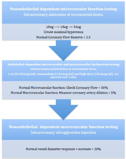
Figure 1: Nonendothelial dependent microvascular function testing.
Non-invasive methods for diagnosis microvascular dysfunction as a reduction in coronary flow reserve include using positron emission tomography [12], cardiac magnetic resonance [24], and transthoracic Doppler echocardiography [14]. We have also introduced these methods into our patient care practices, and use this testing in conjunction to more invasive testing. The use of transthoracic Doppler echocardiography for assessment of measured velocities under hyperemic conditions of adenosine infusion has been used to evaluate for microvascular dysfunction. Using the short-axis view of the left ventricle, diastolic flow can be examined in the anterior groove. Under color Doppler flow mapping, using second harmonic technology, one can identify the distal left anterior descending artery [25]. Bartel et al, found the echocardiographic model comparable to the invasive measurements for microvascular dysfunction assessment [26]. Using an optimal cut off point for coronary flow velocity reserve of 2.84 cm/s they found a sensitivity of 80% and specificity of 88%. This matched previous study data of coronary flow velocity reserve sensitivity and specificity for detection of coronary artery disease [27].
Magnetic resonance myocardial perfusion imaging has been reported to have similar specificity and sensitivity in detecting obstructive CAD compared to conventional radionuclide imaging [24]. Several papers have suggested the utility of Magnetic resonance myocardial perfusion imaging for detection of microvascular dysfunction, however these have been small study populations and sensitivity and specificity data is not available at this time.
Office based testing includes endothelial vasodilator function testing using ENDO-PAT 2000 [28]. This is based on testing done by Gould et al with regards to simulated hyperemia by restriction of maximal flow by temporary occlusion to an artery [22]. Though several methods have been evaluated to diagnose microvascular disease, as mentioned previously the use of intracoronary acetylcholine has proven to be a safe option for evaluation in the cardiac catheterization laboratory. In the follow up to the WISE study, they found in 159 women with chest pain and non-obstructive coronary artery disease, microvascular dysfunction in one-half of the patients on the basis of reduced coronary flow reserve velocities. They found that a coronary reserve velocity of 2.24 provided a 90% sensitivity and 89% specificity for the diagnosis of microvascular dysfunction based on a CFR of < 2.516.
Summary
It is important that we do not overlook the patient population with microvascular dysfunction (especially women), as they have been shown to have higher risk for adverse events and mortality. Through routine testing with coronary angiography bundled with coronary microvascular dysfunction testing, we can give these patients an accurate diagnosis and appropriate management to prevent long-term adverse outcomes. Using CFR testing with a 0.014-inch Doppler coronary wire to determine volumetric flow, any (CFR) velocity measurement of < 2.50 provides a 90% sensitivity and 89% specificity for the diagnosis of microvascular dysfunction16.This protocol has been an important component in the development of our Women’s Heart Disease Program, and plays an important role in the diagnosis of coronary microvascular disease. Ultimately, through routine CFR testing for coronary microvascular dysfunction, we can change management for patients with chest pain syndromes for both symptom relief as well as improved outcomes.
References
- Berne RM. Cardiac nucleotides in hypoxia: possible role in regulation of coronary blood flow. American Journal of Physiology--Legacy Content. 1963; 204: 317-322.
- Patel, Manesh R., Eric D. Peterson, David Dai, J. Matthew Brennan, Rita F. et al. "Low diagnostic yield of elective coronary angiography." New England Journal of Medicine. 2010; 362: 886-895.
- Carpiuc Kimbach T., Deborah L Wingard, Donna Kritz-Silverstein, and Elizabeth Barrett-Connor. "The association of angina pectoris with heart disease mortality among men and women by diabetes status: the Rancho Bernardo Study." Journal of Women's Health. 2010; 19: 1433-1439.
- Sharaf Barry L., Carl J. Pepine, Richard A. Kerensky, Steven E. Reis, Nathaniel Reichek, William J. Rogers, et al. "Detailed angiographic analysis of women with suspected ischemic chest pain (pilot phase data from the NHLBI-sponsored Women’s Ischemia Syndrome Evaluation [WISE] Study Angiographic Core Laboratory)." The American journal of cardiology. 2001; 87: 937-941.
- Johnson, B. Delia, Leslee J. Shaw, Carl J. Pepine, Steven E. Reis, Sheryl F. Kelsey, George Sopko, William J. Rogers et al. "Persistent chest pain predicts cardiovascular events in women without obstructive coronary artery disease: results from the NIH-NHLBI-sponsored Women's Ischaemia Syndrome Evaluation (WISE) study." European heart journal. 2006; 27: 1408-1415.
- Bugiardini, Raffaele, and C. Noel BaireyMerz. "Angina with “normal” coronary arteries: a changing philosophy." Jama. 2005; 293: 477-484.
- Cannon III, Richard O., and Stephen E. Epstein. "“Microvascular angina” as a cause of chest pain with angiographically normal coronary arteries." The American journal of cardiology,1988;61:1338-1343.
- Halcox, Julian PJ, William H. Schenke, Gloria Zalos, Rita Mincemoyer, Abhiram Prasad, Myron A. Waclawiw, et al., "Prognostic value of coronary vascular endothelial dysfunction." Circulation. 2002;106: 653-658.
- Ong Peter, AnastasiosAthanasiadis, Stephan Hill, HolgerVogelsberg, Matthias Voehringer, and UdoSechtem. "Coronary artery spasm as a frequent cause of acute coronary syndrome: The CASPAR (Coronary Artery Spasm in Patients With Acute Coronary Syndrome) Study." Journal of the American College of Cardiology. 2008; 52:523-527.
- Elesber, Ahmad, Amir Lerman, Kevin A. Bybee, Joseph G. Murphy, Gregory Barsness, Mandeep Singh, et al., "Myocardial perfusion in apical ballooning syndrome: correlate of myocardial injury." American heart journal. 2006; 152: e9-13.
- Akashi, Yoshihiro J., David S. Goldstein, Giuseppe Barbaro, and Takashi Ueyama. "Takotsubo cardiomyopathy a new form of acute, reversible heart failure." Circulation. 2008; 118: 2754-2762.
- Drexler H. Factors involved in the maintenance of endothelial function. Am J Cardiol. 1998; 82: 3S-4S.
- Marroquin, Oscar C., Richard Holubkov, Daniel Edmundowicz, Cheryl Rickens, Gerald Pohost, Steven Buchthal, et al. "Heterogeneity of microvascular dysfunction in women with chest pain not attributable to coronary artery disease: implications for clinical practice." American heart journal. 2003; 145: 628-635.
- Lanza, Gaetano A., Antonino Buffon, Alfonso Sestito, Luigi Natale, Gregory A. Sgueglia, Leda Galiuto, et al., "Relation between stress-induced myocardial perfusion defects on cardiovascular magnetic resonance and coronary microvascular dysfunction in patients with cardiac syndrome X." Journal of the American College of Cardiology. 2008; 51: 466-472.
- Sestito, Alfonso, Gaetano A. Lanza, Antonio Di Monaco, Priscilla Lamendola, Giulia Careri, PierpaoloTarzia, "Relation between cardiovascular risk factors and coronary microvascular dysfunction in cardiac syndrome X." Journal of Cardiovascular Medicine. 2011; 12: 322-327.
- Wei Janet, Puja K. Mehta, B. Delia Johnson, Bruce Samuels, SaibalKar, R. David Anderson, et al. "Safety of coronary reactivity testing in women with no obstructive coronary artery disease: results from the NHLBI-sponsored WISE (women's ischemia syndrome evaluation) study." JACC: Cardiovascular Interventions. 2012; 5: 646-653.
- Coffman, Jay D., and Donald E. Gregg. "Reactive hyperemia characteristics of the myocardium." Am J Physiol. 1960; 190: 1143-1149.
- Palmer, R. M., A. G. Ferrige, and S. Moncada. "Nitric oxide release accounts for the biological activity of endothelium-derived relaxing factor." Nature 1987; 327: 524-526.
- Ludmer Paul L., et al. "Paradoxical vasoconstriction induced by acetylcholine in atherosclerotic coronary arteries." New England Journal of Medicine. 1986; 315: 1046-1051.
- Quyyumi AA, Cannon RO III, Panza JA, Diodati JG, Epstein SE. Endothelial dysfunction in patients with chest pain and normal coronary arteries.Circulation. 1992; 86:1864-1871.
- Sueda, Shozo, Akira Oshita, Takahiko Nomoto, YousukeIzoe, Hiroaki Kohno, Hiroshi Fukuda, et al., "Recommendations for performing acetylcholine tests safely: STOP dangerous complications induced by acetylcholine tests (STOP DCIAT)." Journal of cardiology. 2008; 51: 131-134.
- Gould K. Lance, Kirk Lipscomb, and Glen W. Hamilton. "Physiologic basis for assessing critical coronary stenosis: instantaneous flow response and regional distribution during coronary hyperemia as measures of coronary flow reserve." The American journal of cardiology. 1974; 33: 87-94.
- Wilson Robert F., D. E. Laughlin, PETER H. Ackell, W. M. Chilian, M. D. Holida, C. J. Hartley, et al., "Transluminal, subselective measurement of coronary artery blood flow velocity and vasodilator reserve in man." Circulation. 1985; 72: 82-92.
- Doyle Mark, Nicole Weinberg, Gerald M. Pohost, C. Noel BaireyMerz, Leslee J Shaw, George Sopko, et al. "Prognostic Value of Global MR Myocardial Perfusion Imaging in Women With Suspected Myocardial Ischemia and No Obstructive Coronary Disease: Results From the NHLBI–Sponsored WISE (Women's Ischemia Syndrome Evaluation) Study." JACC: Cardiovascular Imaging. 2010; 3: 1030-1036.
- Caiati, Carlo, Cristiana Montaldo, Norma Zedda, Alessandro Bina, and SabinoIliceto. "New Noninvasive Method for Coronary Flow Reserve Assessment Contrast-Enhanced Transthoracic Second Harmonic Echo Doppler." Circulation.1999; 99: 771-778.
- Bartel, Thomas, Ya Yang, Silvana Müller, René R. Wenzel, Dietrich Baumgart, Thomas Philipp, and RaimundErbel. "Noninvasive assessment of microvascular function in arterial hypertension by transthoracic Doppler harmonic echocardiography." Journal of the American College of Cardiology. 2002; 39: 2012-2018.
- Takeuchi, Masaaki, et al. "Assessment of coronary flow velocity with transthoracic Doppler echocardiography during dobutamine stress echocardiography." Journal of the American College of Cardiology. 2001; 38: 117-123.
- Axtell Andrea L., Fatemeh A. Gomari, and John P. Cooke. "Assessing endothelial vasodilator function with the Endo-PAT 2000." Journal of visualized experiments: JoVE’. 2010; 44.