
Case Report
Austin J Clin Case Rep. 2014;1(1): 1005.
Central Retinal Vein Occlusion in Acute Promyelocytic Leukemia and Heterozygosis’ for Factor V Leiden and Methylentetrahydrofolate Reductase (MTHFR) Gene Mutations
Gupta M*, Singh R and Singla N
Department of Medicine, Government Medical College and Hospital, Chandigarh
*Corresponding author: Monica Gupta, Department of Medicine, Government Medical College and Hospital, Address: 1156-C, 32-B, Chandigarh-160030, Chandigarh
Received: May 19, 2014; Accepted: May 27, 2014; Published: May 28, 2014
Abstract
There is a strong link between radiographically detectable coronary calcium and future coronary heart disease events leading to death. Although chest radiographs have low sensitivity for detecting coronary calcification, yet they may be useful in detecting extensive coronary artery disease, especially in the high risk individuals. The chest radiograph provides immense information and is still a useful tool in the resource constraint settings.
Keywords: Coronary artery; Calcification; Chest radiograph
Case Report
A 50-year-old postmenopausal lady was admitted with pyelonephritis to our medical unit. Her past history revealed that she had coronary artery disease for which she underwent coronary artery stenting 2 years back. She also had type 2 diabetes for the past 5 years with good glycemic control. During the medical ward round, interestingly, her chest radiograph revealed a linear calcification of the left anterior descending artery, outlining its course (Figure 1).
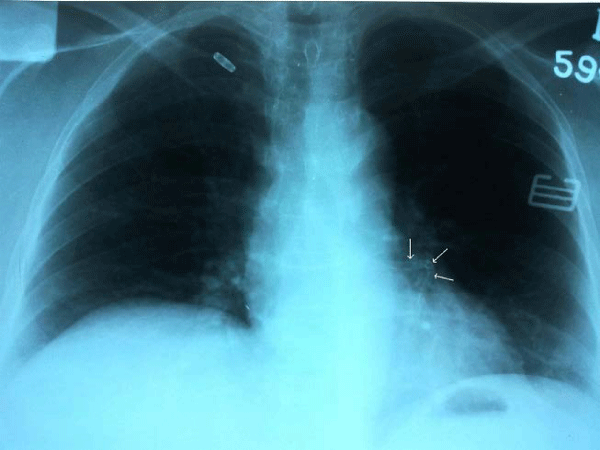
Figure 1 :Patient’s frontal chest radiograph revealing a linear calcification of the left anterior descending artery outlining its course (small white arrows)
Coronary calcification is an active process generally related to widespread atherosclerosis and frequently observed in advanced lesions [1]. There is a strong link between radiographically detectable coronary calcium and future coronary heart disease events leading to death and infarction in symptomatic and very high-risk subjects [2]. Currently, there is an increasing trend to rely on CT detection of coronary calcification to detect subclinical coronary artery disease, but this may not be cost –effective.
Calcifications within the heart, on a chest radiograph, are commonly due to aortic or mitral valve stenosis, prior myocardial infarction resulting a ventricular aneurysm, left atrial wall calcification usually due to rheumatic heart disease or pericardial owing to constrictive pericarditis. All these calcifications have characteristic radiological appearances. Coronary artery stents may be visible on chest X-rays, however these are very small structures and have the radiographic appearance of a tubular structure with a chicken-wire mesh. Especially when the coronaries are heavily calcified, these may not be easily discernable.
Chest radiographs, although readily available and inexpensive, have low sensitivity for detecting coronary calcification [3]. Correlation of such cardiac calcifications with appropriate newer modalities definitely helps to further delineate the anatomical lesions. Fluoroscopy, electron beam, and double-helical computed tomography have enhanced capability to accurately localize coronary calcification and detect smaller/less dense calcific deposits, besides these can also quantify the amount or volume of calcium.
Learning Points
Chest roentgenograms have long been recognized as companions to physicians and complement the diagnostic information. Although it does not detect coronary calcification easily, it may be still affix importance to bedside detection of severe coronary artery disease especially in patients with long standing diabetes. Extensive coronary calcification indicates significant atherosclerotic burden and calls for a more aggressive approach to management of coronary risk factors and an early institution of drug therapy.
References
- Demer LL, Tintut Y. Vascular calcification: Pathobiology of a multifaceted disease. Circulation. 2008; 117: 2938-2948.
- Wexler L, Brundage B, Crouse J, Detrano R, Fuster V, Maddahi J, et al. Coronary artery calcification: Pathophysiology, epidemiology, imaging methods, and clinical implications: Circulation. 1996; 94: 1175-1192
- Kelley MJ, Newell JD. Chest radiography and cardiac fluoroscopy in coronary artery disease. Cardiol Clin. 1983; 1: 575-595.