
Case Report
Austin J Clin Case Rep. 2014;1(4): 1017.
A Case of Giant Fecaloma in a 32-Year-Old Woman
Monica Gupta1*, Purnima Aggarwal2, Ram Singh1 and Lehl SS1
1Department of Medicine, Government Medical College and Hospital, India
2Department of Radiology, Government Medical College and Hospital, India
*Corresponding author: Monica Gupta, Department of Medicine, Government Medical College and Hospital, 1156-C, 32-B, Chandigarh, India
Received: June 02, 2014; Accepted: June 16, 2014; Published: June 18, 2014
Abstract
Chronic constipation is a very common complaint at outpatient clinics. It can progress to fecal impaction if not managed promptly. However rarely it may be extreme causing abdominal distension masquerading an abdominal tumor. We present an unusual case of a 32-year-old mentally challenged female who developed a fecaloma associated with chronic constipation, which was managed conservatively. Diagnosis of fecaloma must be considered in patients presenting with chronic constipation and abdominal mass.
Keywords: Chronic constipation; Fecaloma; Ureters
Introduction
Fecaloma is defined a mass of inspissated feces accumulated in the colon or rectum that is much harder in consistency than impacted feces. The feces initially accumulate, then stagnate and get impacted due to coprostasis, expand and deform the intestine, and develop into large tumor like masses. Fecalomas, often giant ones have been described in Hirschsprung’s disease, psychiatric patients, neglected elderly or bedridden patients, Chagas disease, inflammatory and neoplastic conditions, and in patients suffering from idiopathic chronic constipation.
Clinical Presentation
A 32-year old mentally challenged female reported to our medical unit with 1 year history of progressive constipation and slowly increasing abdominal distension. Caretakers gave the history of 1 bowel movement every third-fourth day with passage of hard stools for initial 6 months, but lately she had spurious diarrhea following daily laxatives use. No history of nausea, vomiting or blood in stools was forthcoming. There was no history of fever, anorexia, weight loss or previous hospitalizations. No urinary complaints were present. Patient had cerebral palsy since childhood and was unable to carry out activities of daily living independently. However there was no history to suggest anorexia or bulimia nervosa.
Examination revealed a normal temperature, supine blood pressure of 110/70 mm Hg, pulse rate of 75/min, a respiratory rate of 14 breaths/min. She weighed 46 kg with good overall nourishment. Abdominal inspection revealed an irregularly distended abdomen especially so in the left half with no evidence of increased peristalsis. During palpation, multiple hard nodular but mobile lumps were palpable in left lumbar, iliac and hypogastric areas. There was no hepatosplenomegaly. Bowel sound appeared normal. Per Rectal examination was reminiscent of rectum loaded with hard feces. No major physical malformations were notable during the examination. Rest of the systemic examination was unremarkable.
Laboratory investigations revealed a hemoglobin 11.2 g/dl, WBC 7000/ mm3, platelets 2.74 lakh/mm and ESR of 24 mm in the first hour. The renal functions and serum electrolytes were normal at creatinine of 0.8 mg/dl, serum Na 140mEq/L, serum K 4.3mEq/L, urinalysis and liver function tests were normal. Chest radiograph was unremarkable. Ultrasound of the abdomen showed evidence of large calcified mass lesions in the para-umblical region extending into the pelvis. Contract enhanced CT abdomen; delayed images revealed dilated rectum as well as sigmoid colon due to loaded fecoliths and extending into the other parts of large bowel (Figure 1a and 1b). Mass effect was also observed on the urinary bladder and bilateral ureters, but there was no evidence of hydronephrosis. Patient was administered repeated enemas, fecal disimpaction per rectum with digital evacuation and laxatives, following which the constipation was partially relieved. Patient did not cooperate for colonoscopy and caretakers refused any further surgical intervention. Patient is under regular follow up for more than 2 years and there has been no complication so far.
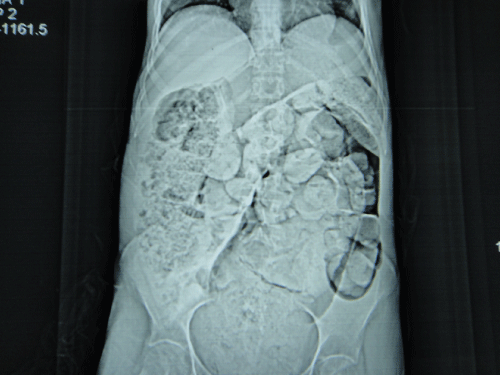
Figure 1a :Coronal image of CECT abdomen showing massively dilated large bowel loaded with fecalomas.
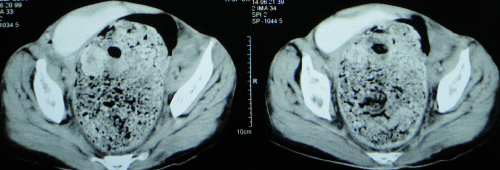
Figure 1b :Axial section of CECT abdomen showing massively dilated rectum with giant fecaloma.
Discussion
Fecaloma is very uncommon condition and an extreme manifestation of fecal impaction, as highlighted above. Symptoms of fecaloma are usually nonspecific with “overflow” type of diarrhea, constipation, unexplained anemia, weight loss and vague abdominal discomfort after meals [1]. Distal colon and rectum are the most common sites for fecalomas [2]. Fecalomas present as abdominal masses mimicking neoplasm’s and can cause local complications by compressing the adjacent anatomical structures, the ureters, urinary bladder, uterus, vagina etc. Occasionally life threatening complications and surgical catastrophes like stercoral perforations, intestinal obstruction, anuria, bladder rupture, peritonitis, and septicemia may occur [3-6].
Most of the fecal impactions are successfully treated by conservative methods such as laxatives, suppositories and trans-rectal enemas. Often it is necessary to dislodge hard stools by manual disimpaction, finger fracture method and digital evacuation. A surgical intervention for uncomplicated fecal impaction is rarely needed [7-9]. Endoscopic approach to removal of fecalomas has also been described [10]. When conservative measures fail or when potentially serious complication (like bowel perforation, peritonitis) supervenes, surgical intervention is essential to prevent mortality. Surgical procedure involves either exploratory laparotomy or laparoscopy followed by removal of fecalomas and resection of the involved colonic segment. It is important to keep a close follow up, prescribe stool softeners and educate the patient about proper dietary habits and involve the patient in regular toilet training sessions.
Conclusion
Fecaloma should be considered in the differential diagnosis of patients presenting with chronic constipation and abdominal mass. Severe chronic constipation should be keenly investigated and should be aggressively approached by appropriate medical, endoscopic and surgical management to prevent significant complications. In high risk patients, treatment is tedious and may necessitate repetitive interventions.
References
- Garisto JD, Campillo L, Edwards E, Harbour M, Ermocilla R. Giant fecaloma in a 12-year-old-boy: a case report. Cases Journal. 2009; 2: 127.
- Rajagopal A, Martin J. Giant Fecaloma with idiopathic sigmoid megacolon: report of a case and review of the literature. Dis Colon Rectum. 2002; 45: 833-835.
- Chute DJ, Cox J, Archer ME, Bready RJ, Reiber K. Spontaneous rupture of urinary bladder associated with massive fecal impaction (fecaloma). Am J Forensic Med Pathol. 2009; 30: 280-283.
- Arana-Arri E, Cortés H, Cabriada V, Lekerika N, García-Verdugo A, Shengelia-Shapiro L. Giant faecaloma causing perforation of the rectum presented as a subcutaneous emphysema, pneumoperitoneum and pneumomediastinum: a case report. Eur J Emerg Med. 2007; 14: 351-353.
- Caiazzo P, De Martino C, Del Vecchio G, Di Lascio P, Marasco M, Laviani F, et al. Megacolon for a giant faecaloma with unlucky outcome: case report and review of the literature. Ann Ital Chir. 2013; 84: 319-322.
- Narang A, Mittal S, Garg P, Aggarwal S, Singh J, Kaushik K, et al. Rectal perforation by impacted fecaloma--a new mechanism proposed. Indian J Gastroenterol. 2013; 32: 417-418.
- Kim KH, Kim YS, Seo GS, Choi CS, Choi SC. A case of fecaloma resulting in the rectosigmoid megacolon. Korean J Gastrointest Motil 2007; 13: 81-85.
- Aiyappan SK, Ranga U, Samraj A, Rajan SC, Veeraiyan S. A case of fecaloma. Indian J Surg. 2013; 75: 323-324.
- Coccolini F, Catena F, Manfredi R, Ansaloni L. A "beehive" in the abdomen. Indian J Surg. 2013; 75: 321.
- Sakai E, Inokuchi Y, Inamori M, Uchiyama T, Iida H, Takahashi H, et al. A: Rectal fecaloma: successful treatment using endoscopic removal. Digestion. 2007; 75: 198.