Case Report
Austin J Clin Case Rep. 2014;1(5): 1022.
Multidetector CT Angiography Images of Vein of Galen Malformation
Amit Nandan Dhar Dwivedi1*, Chandan Mourya1, Ishan Kumar1 and Suchi Tripathi2
1Department of Radiodiagnosis & Imaging, Banaras Hindu University, India
2Department of Internal Medicine, Banaras Hindu University, India
*Corresponding author: Amit Nandan Dhar Dwivedi, Department of Radiodiagnosis & Imaging, Institute of Medical Sciences, Banaras Hindu University, Varanasi 221005, India
Received: May 23, 2014; Accepted: June 30, 2014; Published: July 01, 2014
Abstract
Aneurysm of the vein of Galen is a rare midline arteriovenous malformation seen in children. It makes up approximately 1% of intracranial vascular malformation but 30% of all paediatric vascular malformation. The presentation depends on the age and amount of blood shunted to the malformation. A neonate or a child may present with cardiac failure, macrocephaly, headache, focal neurological signs or subarachnoid haemorrhage. We report a case of a 2 year old male child and rare images that presented with macrocephaly and diagnosed to have vein of Galen aneurysmal malformation on CT scan and CT angiography.
Case Presentation
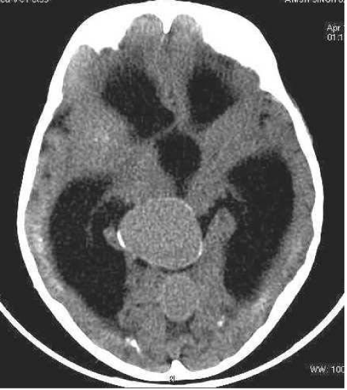
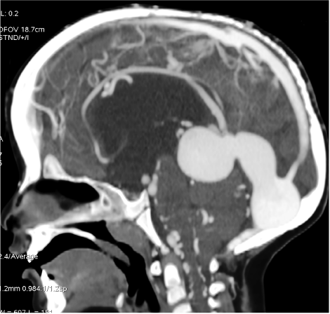
A two year old boy was referred from department of paediatrics to department of radio diagnosis and imaging for evaluation of macrocephaly and delayed development of milestones. The child was born with full term spontaneous vaginal delivery at home. No antenatal ultrasound was performed during this gestation. He had no history of seizure disorder. On systemic examination no significant abnormality was noted. No evidence of any focal neurological deficit was noted. Cardiovascular examination was within normal limits. A non-contrast CT scan [Figure 1] showed a well-defined large rounded homogenous area of increased density with peripheral curvilinear calcification at the posterior third ventricle. There were dilatations of both the lateral and third ventricles. The fourth ventricle was normal. There was no periventricular lucency to suggest acute hydrocephalus. There was no intracranial haemorrhage or midline shift. Contrast examination [Figure 2] showed strong uniform enhancement and confirmed the vascular nature of the lesion and its communication. Diagnosis of vein of Galen aneurysmal malformation was made based on above findings.
Figure 1: Non contrast CT scan showing a well-defined large rounded homogenous area of increased density with peripheral curvilinear calcification at the posterior third ventricle. There were dilatations of both the lateral and third ventricles. The fourth ventricle is normal.
Figure 2: Contrast examination sagittal plane shows robust enhancement and the vascular nature of the lesion.
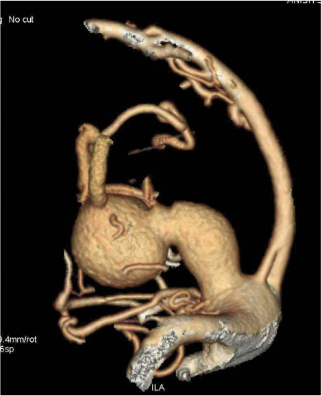
Computerised tomography angiography (CTA) with multi planar reconstruction [Figure 3] showed strongly enhancing rounded lesion at tentorial apex extending posteriorly and draining into dilated superior sagittal sinus through dilated straight sinus. The arterial feeders were noted directly draining into lesion without forming a nidus. Predominant dilated arterial feeders were seen arising from posterior cerebral arteries.
Figure 3: CT angiography with multiplanar reconstruction shows strongly enhancing lesion at tentorial apex extending posteriorly draining into dilated superior sagittal sinus through dilated straight sinus. The arterial feeders were noted directly draining into lesion without forming nidus. Predominant dilated arterial feeders were arising from posterior cerebral arteries.
Discussion
Aneurysm of the vein of Galen is a rare midline arteriovenous malformation seen in children. It makes up approximately 1% of intracranial vascular malformation but 30% of all paediatric vascular malformation. [1,2] The presentation depends on the age and amount of blood shunted to the malformation. A neonate or a child may present with cardiac failure, macrocephaly, headache, focal neurological signs or subarachnoid haemorrhage. [3] In the newborn with a large shunt, severe cardiac failure and cranial bruit are the typical signs. In an infant, an enlarging head due to obstructive hydrocephalus at the aqueduct is a common presentation. In older children, subarachnoid haemorrhage and focal neurological signs may occur. Two methods of classifications had been used extensively in the literature. The Lasjaunias classification is used to classify vein of Galen malformation. It relies upon dividing the entity into choroidal or mural types, depending on the number and origin of feeding arteries. [4] The Yasargil classification is one of the commoner systems for classifying vein of Galen malformation. [5] The diagnosis of vein of Galen aneurysmal malformation can be made using ultrasound Doppler, CT scan, MRI and angiography. The antenatal diagnosis of vein of Galen aneurysm is important to alert the paediatrician of the need for careful postnatal treatment and follow up. Ultrasound is the imaging modality of choice in these cases because of its accuracy, safety, ease of access, low cost and real-time capability. It will usually show an anechoic midline intracranial lesion with blood flow on Doppler. However with the introduction of MRI, especially with echo planar imaging, it can be used for more accurate assessment. MRI has the advantage of better soft tissue signal and contrast and lack of interference by bony structures. CT scan or MRI is important in the early assessment of vein of Galen aneurysm. This is important to assess the brain parenchyma because any evidence of brain damage will contraindicate endovascular treatment. MRI is indeed very important for accurate assessment of the brain parenchyma. The availability of multi detector CT angiography [6] with various options of surface rendering and reformation have aided in the planning of intervention and embolization. Conventional angiography remains the gold standard in the evaluation of the vein of Galen aneurysms. This is only performed if there is plan for endovascular embolization later on. It will show the arterial feeders, the drainage, and indicate whether it is a mural or a choroidal.
Conclusion
Vein of Galen aneurysmal malformation is a rare intracranial vascular malformation commonly seen in the paediatric population. It can be diagnosed in antenatal period by ultra sonography, colour Doppler and MRI. In postnatal period CT and MRI are used to assess brain parenchyma, intracranial bleed & angiography and CT angiography for assessment vascular anomalies for management and classification of malformation.
References
- Raybaud CA, Strother CM, Hald JK. Aneurysms of the vein of Galen: embryonic considerations and anatomical features relating to the pathogenesis of the malformation. Neuroradiology. 1989; 31: 109-128.
- Raybaud CA, Strother CM, Hald JK. Aneurysms of the vein of Galen: embryonic considerations and anatomical features relating to the pathogenesis of the malformation. Neuroradiology. 1989; 31: 109-128.
- Meyers PM, Halbach VV, Dowd CF, Malek AM, Lempert TE, Lefler JE, et al. Hemorrhagic Complications in Vein of Galen Malformations. Ann of Neurol. 2000; 47: 748-755.
- Lasjaunias Pierre L. Vascular diseases in neonates, infants and children. Springer. 1997.
- Yasargil MG. Microneurosurgery IIIB. New York: Thieme Medical Publishers. 1988. pp. 323-357.
- Martelli A, Scotti G, Harwood-Nash DC, Fitz CR, SH Chuang. Aneurysms of the Vein of Galen inChildren : CT and Angiographic Correlations. Neuroradiology. 1980; 20: 123-133.


