Clinical Image
Austin J Clin Case Rep. 2014;1(7): 1031.
Optic Disc Pit in a Young Female Patient
Panos GD*
Department of Ophthalmology, University of Geneva, Switzerland
*Corresponding author: Georgios Panos, Department of Ophthalmology, University of Geneva, Geneva University Hospitals, Alcide-Jentzer 22, CH 1211, Geneva, Switzerland
Received: June 30, 2014; Accepted: July 22, 2014; Published: July 25, 2014
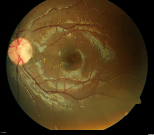
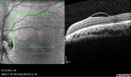
An 8 year old girl, with clear family history, has been addressed to our Paediatric Ophthalmology & Neuro-ophthalmology Unit for an optic neuropathy suspicion in her left eye (OS). The clinical examination revealed a hypertelorism with exotropia OS. The visual acuity (VA) was 20/20 right eye (OD) and 20/35 OS. The cycloplegic refraction was +0.50 -0.25 X 160 OD and +0.75 -0.25 X 165 OS. Goldmann applanation tonometry was 14mm Hg for both eyes. Anterior segment examination was within normal limits for both eyes. Dilated fundus examination revealed an optic disc pit (ODP) on the temporal side of the optic disc with possible vitreomacular traction OS (Figure 1, yellow arrows). Optical Coherence Tomography (OCT) verified the vitreomacular traction (Figure 2).
Figure 1 :
Figure 2 :
ODPs are extremely rare, with an incidence of 1:11,000 for congenital ODPs, and are frequently associated with maculopathy (serous retinal detachment, cystoid macular oedema, vitreomacular traction) [1-3]. Surgical approach involving pars plana vitrectomy is a widely accepted treatment with promising results [4,5].
References
- Chang M. Pits and crater-like holes of the optic disc. Ophthalmic Semin. 1976; 1: 21-61.
- Kranenburg EW. Crater-like holes in the optic disc and central serous retinopathy. Arch Ophthalmol. 1960; 64: 912-924.
- Theodossiadis PG, Grigoropoulos VG, Emfietzoglou J, Theodossiadis GP. Vitreous findings in optic disc pit maculopathy based on optical coherence tomography. Graefes Arch Clin Exp Ophthalmol. 2007; 245: 1311-1318.
- Dai S, Polkinghorne P. Peeling the internal limiting membrane in serous macular detachment associated with congenital optic disc pit. Clin Experiment Ophthalmol. 2003; 31: 272-275.
- Ghosh YK, Banerjee S, Konstantinidis A, Athanasiadis I, Kirkby GR, Tyagi AK, et al. Surgical management of optic disc pit associated maculopathy. Eur J Ophthalmol. 2008; 18: 142-146.

