
Case Report
Austin J Clin Case Rep. 2014;1(7): 1032.
Metastasis to Bartholin Gland: An Extremely Rare Presentation of Relapse from Endometrioid Adenocarcinoma of Uterus
Pushpalatha K1*, Sharma DN2, Kumar S1 and Kumar R3
1Department of Obstetrics and Gynaecology, All India Institute of Medical Sciences, India
2Department of Radiation Oncology, All India Institute of Medical Sciences, India
3Department of Nuclear Medicine, All India Institute of Medical Sciences, India
*Corresponding author: Pushpalatha, Department of Obstetrics and Gynaecology, All India Institute of Medical Sciences (AIIMS), Near BijuPatnaik Police Academy, Village Sijua, Bhubaneswar - 757019, Odisha, India
Received: June 28, 2014; Accepted: July 21, 2014; Published: July 25, 2014
Introduction
Endometrial cancer is the most common cancer of the female reproductive tract and the fourth most common cancer overall in women, accounting for an estimated 43,470 new cases and 7,950 deaths in the United States in 2010. [1] Recurrent and advanced endometrial cancer remains a treatment dilemma. However, patients diagnosed with advanced disease continue to have a dismal prognosis with a 17% 5-year survival [2]. Concomitant Bartholin gland and Brain metastases from endometrial carcinoma are rare. The incidence of brain metastases from endometrial carcinoma ranges from 0.3% to 1.4%. [3,4]. Here we report a rare case of endometrial carcinoma with such metastases.
Case Presentation
A 62-year-old multiparous postmenopausal woman was referred to our Gynae-oncology clinic as a postoperative case of Carcinoma Endometrium (FIGO stage IIIC grade II) for adjuvant radiotherapy and chemotherapy. She had elsewhere under gone laparotomy with extra fascial hysterectomy, bilateral Salpingo-oophorectomy, bilateral pelvic lymphadenectomy, infracolic omentectomy on April 25, 2009. Histopathological report showed moderately differentiated endometrioid adenocarcinoma infiltrating more than half thickness of the myometrium. Ovary was involved by poorly differentiated adenocarcinoma. Among 7 pelvic lymph nodes, 1 obturator lymph node was involved by the tumour. Omentum and bilateral parametria were free. Patient received external beam radiotherapy of 50.4 Gy/28#/5.5 weeks W.E.F 5th June till 7th August 2009 followed by 2 sessions of intra vaginal brachy therapy till 18th August 2009. 6 cycles of hemotherapy comprising of Paclitaxel (260 mg IV/D1) and Carboplatin (450 mg IV/D2) was administered at an interval of 3 weeks from Jan 2010-April 2010. After an interval of 6 months, patient developed pruritus vulva associated with left Bartholin’s gland swelling. Vulval examination revealed 3x3cms left Bartholin solid mass with smooth surface without any discharge. There were no grossly enlarged inguinal lymph nodes bilaterally. Per speculum examination revealed normal vault. Biopsy of the Bartholin lesion showed adenocarcinoma. CT, PET and PET-CT axial section and whole body projection images (Figure1) showed focal area of abnormal 18-Fluoro Deoxy Glucose (FDG) uptake in the region of left Bartholin gland and mild FDG uptake in left inguinal lymph nodes suggestive of metastasis. Concurrent CECT brain done for headache also revealed multiple brain metastases. In view of advanced disease, patient was administered palliative WBRT 20 Gray in 5 fractions between 13-10-10 to 19-10-10. Patient was seen last on Dec 2011 with progressive brain metastasis, left hemiplegia as well as local progression of the disease. She ultimately succumbed to death due to progressive disease on Feb 2012 making the overall survival of 2 years and 10 months.
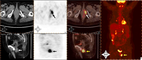
Figure 1 : Showed focal area of abnormal FDG uptake in the region of left Bartholin gland and mild FDG uptake in left inguinal lymph nodes suggestive of metastasis.
Discussion
Generally endometrial cancer carries a good prognosis. However, metastatic tumors of the vulva especially to Bartholin gland are rare. Moreover, in the presence of metastatic endometrial carcinoma to the vulva, it is necessary to verify if other visceral metastases are present. [5] Metastasis from an endometrial carcinoma may occur to adjacent organs by direct extension or in the form of distant metastasis (lungs, liver or bones) by lymphatic or hematogenous spread. Skin metastasis may also occur by direct extension, lymphatic dissemination or by implantation at the site of surgical scars.
Dehner in 1973, after analyzing the different forms of dissemination of endometrial tumors, stated that the fact that vascular involvement is common tends to confirm that this is the mode of dissemination to the vulva and the route of distant metastasis. However, the possibility of lymphatic dissemination cannot be discarded. With respect to the vulva as a site of metastasis, the author found 8.4% of metastatic tumors of the vulva in a series of 262 cases of malignant tumors at this site [6]. Between 1944 and 2001, Neto et al. in 2003 reported 66 cases of vulval metastasis; out of which, six of these involved endometrial carcinoma. The majority of metastatic tumors consisted of nodules and involved the labia majora. Only one case involved the clitoris. In 87% of cases, the patient died at a mean of 7.5 months after diagnosis, reflecting the fact that this is normally a terminal condition. [7] Filho et al (2014) have reported another case of clitoral metastasis from endometrial carcinoma who died 6 months after diagnosis of metastatic lesion following treatment with palliative radiotherapy [8]. Giordano et al (2005) reported a case of hepatic and vulval metastasis in the form of a plaque on the commissure of the labia majora 8 months after hysterectomy for a grade 3 endometrioid adenocarcinoma. The same authors recommend investigating distant metastasis in visceral organs following diagnosis of vulval metastasis from an endometrial carcinoma [9].
In our case, concurrent brain metastasis was also found. However disseminated metastasis led to progressive left hemiplegia along with local disease progression making the disease not amenable to treatment. In large series, endometrial carcinoma comprises approximately 1% of all central nervous system (CNS) metastases, and it is only the third most common female genitourinary malignancy to involve the brain, behind ovarian carcinoma and choriocarcinoma. However, its incidence may be increasing secondary to improved diagnosis, more effective management of the primary disease, and overall increased survival. Brain metastases from endometrial carcinoma tend to occur in the context of widely disseminated disease. Correspondingly, the prognosis of patients with brain metastases is historically poor, with a median survival of 1 to 2 months. In our case, patient survived the metastatic disease for 1 year 4 months making the overall survival of 2 years and 10 months. Current recommendations support the use of aggressive multimodality treatment, including a combination of surgical resection or SRS (Stereotactic radiosurgery) with WBRT for brain metastases from endometrial carcinoma [10].
Possibility of double primary is considered when the histopathology is often different. Interestingly median age of occurrence (50 years) and histology (adenocarcinoma) are similar in endometrial carcinoma and Bartholin gland carcinoma. Bartholin gland carcinomas are slow-growing tumours associated with frequent recurrences that exhibit local invasion, and metastasis to tissues and/ or organs. Bones and the lungs are the most common sites of distant recurrence [11-14]. As far as treatment of Bartholin Gland Carcinoma is concerned, certain investigators hypothesize that radiation therapy or chemoradiation offer effective alternative strategies to surgery, whilst preserving genital function and maintaining low levels of morbidity [11-13]. Early diagnosis combined with a radical vulvectomy and bilateral inguinal femoral lymph node dissection may optimize the patient’s likelihood of survival [11,15]. Patients that do not exhibit metastatic lesions at early diagnosis should undergo cancer lesion excision to reduce the tumour payload and increase the efficacy of radiotherapy. In the present case, biopsy from Bartholin gland swelling revealed adenocarcinoma consistent with that of primary endometrial adenocarcinoma. As patient had concurrent brain metastasis, whole brain radiotherapy and chemotherapy was offered as a multimodality treatment.
Conclusion
Bartholin gland metastasis with concurrent distant metastasis from an endometrial carcinoma has a poor prognosis and rapid and aggressive progression. Little is known on the association between endometrial cancer and metastasis to the Bartholin gland. Hence it is necessary to emphasize the importance of early diagnosis and intervention, and raising awareness about regular gynaecological exam in women to take care of their health. Knowledge of this rare metastatic site will help the interpreting physician make the correct diagnosis and also warrants to search for concurrent distant metastasis for appropriate management.
References
- Jemal A, Siegel R, Xu J, Ward E. Cancer statistics, 2010. CA Cancer J Clin. 2010; 60: 277-300.
- Dizon DS, Blessing JA, McMeekin DS, Sharma SK, Disilvestro P, Alvarez RD. Phase II trial of ixabepilone as second-line treatment in advanced endometrial cancer: gynecologic oncology group trial 129-P. J Clin Oncol. 2009; 27: 3104-3108.
- Cormio G, Lissoni A, Losa G, Zanetta G, Pellegrino A, Mangioni C. Brain metastases from endometrial carcinoma. Gynecol Oncol. 1996; 61: 40-43.
- Orrru S, Lay G, Dessi M, Murtas R, Deidda MA, Amichetti M. Brain metastases from endometrial carcinoma: report of 3 cases and review of the literature. Tumori. 2007; 93: 112-117.
- Fakor F, Hajizadeh Falah H, Khajeh Jahromi S. Endometrial carcinoma metastatic to the clitoris: a case report and review of the literature. Acta Med Iran. 2013; 51: 652-654.
- Dehner LP. Metastatic and secondary tumors of the vulva. Obstet Gynecol. 1973; 42: 47-57.
- Neto AG, Deavers MT, Silva EG, Malpica A. Metastatic tumors of the vulva: a clinicopathologic study of 66 cases. Am J Surg Pathol. 2003; 27: 799-804.
- Filho AC, Garbeloto E, Santiago KC, da Motta LL. Endometrial carcinoma metastatic to the clitoris: A case report. Gynecol Oncol Case Rep. 2014; 8: 1-3.
- Giordano G, Gnetti L, Melpignano M. Endometrial carcinoma metastatic to the vulva: a case report and review of the literature. Pathol Res Pract. 2005; 201: 751-756.
- Monaco E, Kondziolka D, Mongia S, Niranjan A, Flickinger JC, Lunsford LD. Management of brain metastases from ovarian and endometrial carcinoma with stereotactic radiosurgery. Cancer. 2008; 113: 2610-2614.
- Zhan P, Li G, Liu B, Mao XG. Bartholin gland carcinoma: A case report. Oncol Lett. 2014; 8: 849-851.
- Massad LS, De Geest K. Multimodality therapy for carcinoma of the Bartholin gland. Gynecol Oncol. 1999; 75: 305-307.
- López-Varela E, Oliva E, McIntyre JF, Fuller AF Jr. Primary treatment of Bartholin's gland carcinoma with radiation and chemoradiation: a report on ten consecutive cases. Int J Gynecol Cancer. 2007; 17: 661-667.
- Kumar R, Singhal M, Acharya R, Chawla N. Adenoid cystic carcinoma of Bartholin’s gland - A rare entity likely to be misdiagnosed. Rev EspPatol. 2011; 44: 213–215. (In Spanish)
- Hwang TL, Hung YC, Chang HW. Adenoid cystic carcinoma of Bartholin's gland. Taiwan J Obstet Gynecol. 2012; 51: 119-120.