
Case Report
Austin J Clin Case Rep. 2015;2(2): 1070.
Milky Plasma
Sharon Leung*
Division of Critical Care Medicine, Department of Medicine, Albert Einstein College of Medicine, USA
*Corresponding author: Sharon Leung, Division of Critical Care Medicine, Albert Einstein College of Medicine, New York.
Received: December 02, 2014; Accepted: April 13, 2015; Published: April 20, 2015
Case Report
A 27-year-old woman with a history of diabetes mellitus and hypertriglyceridemia-induced pancreatitis presented with acute onset of severe constant mid-epigastic pain radiating to the back. On admission, an abdominal exam was notable for epigastic tenderness and mild distention. Lower extremities revealed hypertrophied calves. Initial laboratory data were significant, with sodium 126 meq/L; ionized calcium 3.13 mg/dl; hematocrit 35.6%; triglycerides 10,658 mg/dl and lipase 420 U/L. Computed tomography demonstrated acute severe necrotizing pancreatitis involving neck and tail. Over the next eight hours, eight liters of crystalloid continuous insulin drip was administered with aggressive electrolyte replacement. Repeat laboratory data showed sodium 120meq/L; ionized calcium 2.89 mg/dl; lipase 1,634 U/L; hematocrit 42.7% and creatinine 0.5mg/dl. Emergent apheresis with fresh frozen plasma was performed; (Figure 1) shows the plasma of the patient. Post apheresis, laboratory data showed triglyceride 1,603 mg/dl; hematocrit 31.9% and sodium 137meq/L. Six hours later, the patient developed severe abdominal compartment syndrome (intra-abdominal pressure 46 mmHg) with acute anuria. She underwent emergent laparostomy. On postoperative day 13, she developed pulmonary embolism with bilateral lower extremities deep vein thrombosis. The patient subsequently recovered and was discharged home on day 18.
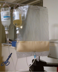
Figure 1: Milky Plasma.