
Special Article - Cancer Clinical Cases and Images
Austin J Clin Case Rep. 2015; 2(4): 1080.
Primary Malignant Melanoma of Esophagus
Dianbo Cao*
Department of Radiology, The First Hospital of Jilin University, China
*Corresponding author: Dianbo Cao, Department of Radiology, The First Hospital of Jilin University, China
Received: October 13, 2015; Accepted: November 04, 2015; Published: November 06, 2015
Case Presentation
A 63-years old woman presented with progressive dysphagia for 1 month, and had lost approximately 2kg in weight since the onset of his illness. Physical examination showed no abnormality. MSCT scan revealed a solid mass in the distal third of the esophagus, where local fat space around the esophagus still remained (Figure A, B). Endoscopy revealed an irregular dark endoluminal and lobulated mass suited 30-35cm from the incisors (Figure C). Endo sonography showed esophageal mass to be solid, heterogenous in echoarchitecture and arising from the deep mucosa (Figure D). The biopsy specimen suggested suspected malignant melanoma. Extensive detailed examination revealed no other skin, anal, facial or rectal lesions. The patient underwent an Ivor-Lewis esophagogastrectomy and lymph node dissection. The surgical specimen showed a 5.0 cm ×4.0 cm ×4.0cm, polypoid and pigmented lesion located in the distal esophagus. The tumor was composed of plump spindle-shaped cells with pigmented cytoplasm, prominent nuclei and numerous mitosis (Figure E). Immunohistochemical stains were positive for S-100 protein and HMB-45(Figure F). Therefore, a diagnosis of primary malignant melanoma of esophagus (PMME) was made. The patient had an uneventful recovery and was in good condition on the followup of 1 year.
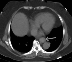
Figure 1A:
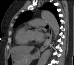
Figure 2B:
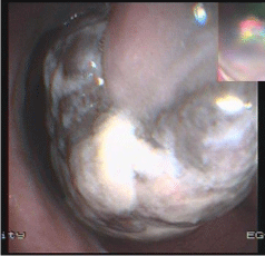
Figure 3C:
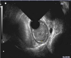
Figure 4D:
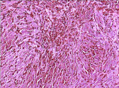
Figure 5E:

Figure 6F:
Malignant melanoma is a typical cutaneous tumor, originating from melanocytes. However, the origin of melanocytes in the esophagus remains hypothetical. PMME is exceedingly rare, varying its incidence from 0.1% to 0.2% of all esophageal malignant tumors [1]. PMME may be found in all areas of the esophagus, but has predominantly been located in the lower two-thirds of the esophagus. PMME occurs twice as frequently in men as in women and usually presents during the sixth and seventh decades of life. Imaging studies such as barium meal and CT only reveal esophageal occupying lesion. The characteristic appearances is a polypoid irregular pigmented and friable lesion at endoscopy, but only 54.7% of cases are diagnosed preoperatively as malignant melanoma. Endoscopic ultrasound is seldom applied and common a hypoechoic or a mixed echogenicity mass, as described in our case. Because of the predominant submucosal location of the tumor in more than 50% of the cases, endoscopic biopsies were negative or misinterpreted as poorly differentiated squamous cell carcinoma [2].
PMME is a rare disease that is characterized by aggressive invasion, early metastasis and poor prognosis [3]. Treatment protocols are not well-established, but a esophagectomy is still the treatment of choice.
References
- Kelly J, Leader M, Broe P. Primary malignant melanoma of the oesophagus: a case report. J Med Case Reports. 2007; 14: 50.
- Ahlawat SK, AL-Kawas FH, Ozdemirli M. Esophageal polypoid mass in a 78-year-old woman. Gastroenterology. 2011; 140: 405,737.
- Yu H, Huang XY and LiY . Primary malignant melanoma of the oesophagus: a study of clinical features, pathology, management and prognosis. Dis Esophagus. 2011; 24: 109-113.