
Case Report
Austin J Clin Case Rep. 2016; 3(4): 1099.
An Unusual Case of Rectal Metastasis from Ovarian Cancer
Slimani KA¹*, Debbagh A¹, Torreis M¹, Sbitti Y¹, Errihani H² and Ichou M¹
¹Department of Medical Oncology, Military Hospital Mohammed V, Rabat, Morocco
²Department of Medical Oncology, National Institute of Oncology, Rabat, Morocco
*Corresponding author: Alaoui Slimani K, Department of Medical Oncology, Military Hospital Mohammed V, Rabat, Morocco
Received: July 21, 2016; Accepted: September 13, 2016; Published: September 16, 2016
Abstract
Rectal metastasis is a rare localization from ovarian cancer. We report a case of ovarian adenocarcinoma that had rectal metastases at the releapse, diagnosed by cytomorphological features and immunocytochemical staining.
Keywords: Rectal metastasis, Ovarian carcinoma, Immunohistochemistry
Abbreviations
AUC: Area Under Curve; CT: Computed Tomography; CMT: Chemotherapy
Introduction
Primary ovarian cancers tend to spread, at first, within the peritoneal cavity and the omentum. Colorectal metastasis from primary ovarian carcinoma account for approximately 4% and an isolated rectal metastasis are very rare [1,2].
Case Presentation
We report a 70-year-old female which was operated for ovarian sero-papillary adenocarcinoma in 2005 for which she received a total hysterectomy and bilateral oophorectomy without lymphadenectomy or adjuvant therapy. In 2009, she presented an isolated centropelvic recurrence and she received six cycles of chemotherapy based on paclitaxel (175 mg/m²) - carboplatin (AUC6) regimen, then surgery completed by three cycles of carboplatin alone because of persistant neuropathy. In 2014, she presented a persistant diarrhea, colonoscopy objectified a rectal tumor, a biopsy revealed rectal metastasis of ovarian carcinoma; Immunohistochemical staining was positive for cytokeratin 7 and negative for cytokeratin 20 (Figures 1 & 2). Computed tomography (CT) scan showed lung micrometastasis and abdominal lumph node metastasis. She received 6 cycles of chemotherapy based on gemcitabine (1000 mg/m²) carboplatine (AUC6) regimen, the evaluation showed a partial response. Three months later, she progressed clinically and radiologically, then, she received metronomic cyclophosphamide (50 mg/day) with good tolerance. Currently, she is stable under surveillance.
Discussion
Rectal metastasis from ovarian cancer is very rare. Koyama et al. have reported only 19 such cases since 2005 in Japan [3]. Therefore, distinguishing rectal metastasis from ovarian carcinoma and primary rectal cancer based on the macroscopic appearance is difficult. Immunohistological staining is very useful to differentiate the origin, Loy et al. reported a cytokeratin 7 positive/cytokeratin 20 negative immunophenotype to be nearly 100% specific for an ovarian origin [4]. In our case, immunohistological staining is consistent with an ovarian origin.
The most plausible explanation of colorectal involvement in ovarian adenocarcinomas is through intra-peritoneal seedling [5].
Identification of the correct primary tumor is necessary for an optimal management, including, specific Chemotherapy (CMT) in advanced stages. Because ovarian adenocarcinomas respond to platinum based CMT, and rectal adenocarcinomas respond to 5-fluorourocil based CMT [6].
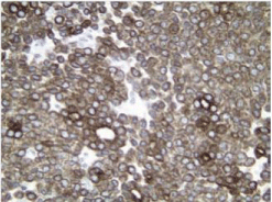
    Figure 1:  Positive immunohistochemical staining for cytokeratin 7
(Immunostain Cytokeratin 7 x 40).
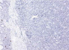
    Figure 2:  Negative immunohistochemical staining for cytokeratin 20
(Immunostain Cytokeratin 20 x 40).
For the treatment, if we have localized rectal metastasis, we should discuss metastasectomy. O’Hanlan et al. have reported that a bowel resection with a wedge resection of mesentery, including paracolic and intermediate-level nodes might be indicated to achieve optimal debulking of gastrointestinal metastases from ovarian carcinomas [7]. But in our case, there were other metastases. Palliative chemotherapy is indicated using platinium based regimen.
Conclusion
It is important to differentiate primary and metastasis rectal carcinomas, because prognosis and treatment differ significantly. Immunohistochemistry can be helpful in solving these dilemmas.
References
- Nakao Y, Suzumura K, Nagata H, Daiwa Y, Arikawa S, Ando K. A case of colon metastasis of ovarian cancer. J Chubu Surg Soc. 2005; 41: 92.
- Haraoka S, Iwashota A, Nakayama Y. Metastatic tumor of the gastrointestinal tract. Stomach Intestine. 2003; 38: 1755-1771.
- Kohyama M, Takesue Y, Ohge H, Sakashita M, Murakami Y, Sueda T. A case of colon metastasis from ovarian cancer presented with intussusception. J Jpn Surg Assoc. 2005; 66: 2767 71.
- Loy TS, Calaluce RD, Keeney GL. Cytokeratin immunostaining in differentiating primary ovarian carcinoma from metastatic colonic adenocarcinoma. Mod Pathol. 1996; 9: 1040-1044.
- Tei S, Murata T, Sibuya M, Shibutani M, Yamada S, Kanemura S. A case of colon metastasis of ovarian cancer. J Jpn Surg Assoc. 2005; 66: 886-888.
- Sobrero AF, Aschele C, Bertino JR. Fluorouracil in colorectal cancer - a tale of two drugs: implications for biochemical modulation. J Clin Oncol. 1997; 15: 368-381.
- O’Hanlan KA, Kargas S, Schreiber M, Burrs D, Mallipeddi P, Longacre T, et al. Ovarian carcinoma metastases to gastrointestinal tract appear to spread like colon carcinoma: implications for surgical resection. Gynecol Oncol. 1995; 59: 200-206.