
Case Report
Austin J Clin Case Rep. 2016; 3(5): 1106.
Unilateral Mydriasis during Orbital Floor Fracture Reconstruction: A Case Report
Angelo DF* and Santos R
Department of Stomatology, University of Lisbon, Portugal
*Corresponding author: David S. Faustino Angelo, Department of Stomatology, Santa Maria Hospital, University of Lisbon, Portugal
Received: September 19, 2016; Accepted: November 30, 2016; Published: December 20, 2016
Abstract
Introduction: Orbital floor fractures are the most common traumatic fractures in the facial skeleton. The occurrence of mydriasis during orbital floor fracture reconstruction may cause significant distress to the surgeons, as it may be associated with potential severe complications.
Case Presentation: A nineteen years old man presented a left medial orbital wall and orbital floor fracture. Ocular injury was ruled out by Ophthalmology and surgical approach was planned.
Management and Outcome: The surgical approach consisted in transconjuntival incision and internal fixation with titanium mesh. After mesh positioning, left eye mydriasis was detected. It persisted for three hours, reverting spontaneously afterwards. Therefore, operative vasoconstrition with lidocaine and epinephrine was considered to be the cause of left mydriasis.
Discussion: Detailed examination of both eyes preoperatively highly reduces anxiety throughout possible complications. Indeed, surgeon must be aware of lidocaine and epinephrine side effects in iris muscular complex � mydriasis.
Keywords: Mydriasis; Orbital floor fracture; Lidocaine; Epinephrine
Introduction
Orbital floor fractures are the most common traumatic fractures in the facial skeleton, being the orbital floor and medial wall commonly affected [1,2]. The occurrence of mydriasis during repair of an orbital fracture can be a distressing event. Indeed, previous studies reported 2.1% incidence of intraoperative mydriasis [3]. Regarding its causes, they are resumed in Table 1. Finally, the combination of careful preoperative evaluation and planning, as well as specific intraoperative investigations when mydriasis is encountered, can be immensely valuable in assuaging surgeons’ anxiety during this surgical procedure. We report a case of intra operative mydriasis due to local anesthetic infiltration.
Case Presentation
A nineteen years old patient with no relevant medical or familiar history, presented with an orbital trauma. The patient referred conserved visual acuity without diplopia. Detailed examination of both eyes, including ocular movement, assessment of pupillary size, direct pupillary light reflex, indirect ophthalmoscopy and screening for enophthalmos was performed. Computed tomography revealed a left medial orbital wall and orbital floor fracture without inferior rectus entrapment.
Management and Outcome
Surgical technique consisted in orbital vasoconstrition (subconjunctival administration of 1.8 ml of lidocaine with epinephrine 2%-1:80000). Conjunctival flap was pulled up to be sutured to the upper lid for corneal protection which unable the surgeon to control pupil variation during surgery. After exposing the left medial orbital wall and orbital floor fracture, it was measured the defect size under optic nerve visualization. The titanium mesh was cut to adapt in the medial wall and orbital floor. Under adequate retraction of the intraorbital soft tissues, the mesh was positioned in order to achieve a stable recontouring of the medial wall and orbital floor. After reconstrution, conjuctival flap was released and left mydriasis was detected (Figure 1). Anesthetic drug assessment revealed fentanyl as the only drug directly involved in pupillary size and reflexes. It causes miosis, and thereby, cannot be responsible for the pupillary finding. However, subconjuntival lidocaine, as well as epinephrine, may cause unilateral mydriasis. The surgical team assumed that the previously performed orbital vasoconstriction was the main cause of the unilateral mydriasis and the surgery pursued. The patient’s mydriasis persisted for three hours hereupon it reverted spontaneously. The postoperative course was uneventful and no visual changes occurred until ward’s discharge. After three months, the patient revealed a good visual outcome with no deficit observed on clinical examination (Figure 2).
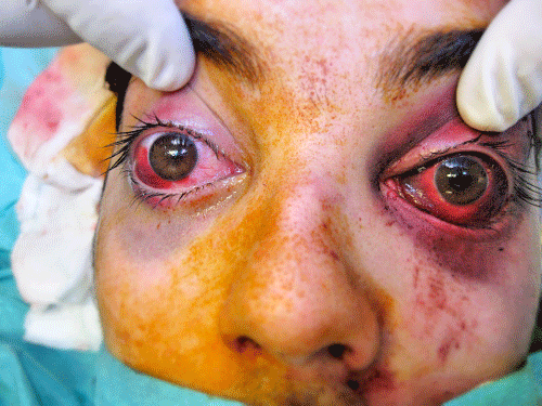
Figure 1: Intraoperative left mydriasis.

Figure 2: Three months after surgical approach.
Discussion
This case report emphasizes the importance of careful pupil examination prior to surgery. Indeed, alterations in the pupillary size can occur intraoperatively due to a great variety of factors. For instance, the use of narcotics (e.g., fentanyl) is associated with nonreactive and miotic pupils [1]. On the other hand, the injection of an anesthetic mixture containing lidocaine and epinephrine into conjunctiva or eyelids can lead to its diffusion into the iris muscular complex, leading to cycloplegia and consequent mydriasis [3]. The most common causes of intraoperative mydriasis are local anesthetic diffusion (solutions containing epinephrine) and traction of inferior rectus and oblique muscles causing temporary neuropraxia of the oculomotor nerve inferior branch [1]. This phenomenon appears to be more common with the transconjunctival approach (2.1%, anesthetic injected into lower conjunctiva) than the subciliary approach (0.8%, anesthetic injected into lower eyelid skin) 3. Direct iatrogenic injury to the optic nerve is less likely to occur. The optic canal is located approximately 45 mm posterior to the infraorbital rim and iatrogenic damage to the optic nerve is an uncommon occurrence in experienced hands [4,5]. Should this occur, it may be detected by the presence of intraoperative relative afferent pupillary defect. Frequently, this is difficult to evaluate due to the concomitant use of intravenous narcotics, which decrease the afferent and efferent visual pathway [1]. Therefore, preoperative detailed examination of both eyes is mandatory. If mydriasis is observed and intraoperatively vasoconstriction was performed, one can assume that local anesthetic and epinephrine diffusion is the main responsible for the event and surgery must proceed with careful postoperative assessment to exclude other possible causes.
![]()
    1.    Systemic causes


    1.1. Pharmacological    agents: atropine, ephedrine, epinephrine, norepinephrine.


    1.2. Evolution    of closed intracranial events: intracranial hemorrhage, rupture of    intracranial aneurysm, cerebral anoxia.


    2.    Ocular causes


    2.1. Local    anesthetic infiltration-eyelid, orbit, paranasal sinuses (cocaine).


    2.2.    Orbital complications-manipulation of inferior division of III nerve,    manipulation of the ciliary ganglion, direct injury to the optic nerve.


    2.3. Undiagnosed    open globe injury with intraoperative rupture.


    2.4. Violation    of poorly repaired/healed open globe with intraoperative dehiscence.
Table 1: Intraoperative causes of mydriasis [1]. Mydriasis during Orbital Floor Fracture Reconstruction.
References
- Matthew S, Al-Mousa R, Sundar G, Lim TC. Mydriasis during orbital floor fracture reconstruction: a novel diagnostic and treatment algorithm. Craniomaxillofac Trauma Reconstruction. 2010; 3: 209-216.
- Gonzalez MO, Durairaj VD. Indirect orbital floor fractures: A meta-analysis. Middle East Afr J Ophthalmol. 2010; 17: 138-141.
- Lim TC, Tan WT, Lim J, Cheng LC. Where’s the point? Plast Reconstr Surg. 1998; 101: 861-862.
- Lemke BN, Lucarelli MJ. Anatomy of ocular adnexa and orbit. Ed. Smith BC, In: Ophthalmic Plastic and Reconstructive Surgery. 2nd ed. St. Louis, MO: CV Mosby. 1997: 3-78.
- Whitnall SE. The Anatomy of the Human Orbit and Accessory Organs of Vision. New York: Oxford University Press. 1932; 1-252.