
Case Report
Austin J Clin Case Rep. 2017; 4(1): 1112.
Utility of Dual-Energy Computed Tomography for Evaluation of Silicone within Internal Mammary Nodes
Doerge S, Glazebrook KN*, Leng S and McCollough C
Department of Radiology, Mayo Clinic, USA
*Corresponding author: Glazebrook KN, Department of Radiology, Mayo Clinic 200 First Street SW, Rochester, MN 55905, USA
Received: October 19, 2016; Accepted: January 11, 2017; Published: January 31, 2017
Abstract
Silicone implants are commonly used for breast reconstruction following breast cancer surgery. Over time the silicone envelope weakens and may rupture. The free silicone may remain within the fibrous capsule (intracapsular rupture), extend into the adjacent breast tissue (extracapsular rupture) or may be taken up by macrophages and can be found in the regional nodes. It is well recognized to be deposited in axillary lymph nodes however there have been few case reports of internal mammary silicone adenopathy. Silicone granulomata within axillary and internal mammary nodes can have high tracer uptake on F-18 FDG positron emission Tomographic Computed tomography (PET/CT), mimicking breast cancer. Dual-energyCT (DECT) allows determination of the density and atomic number of tissue thereby providing material composition information. We present a case of silicone axillary and internal mammary lymphadenopathy mimicking recurrent breast cancer on PET/CT, but with silicone clearly identified on DECT.
Keywords: Dual energy computed tomography; Silicone implants; Breast cancer
Abbreviations
CT: Computed Tomography; DC: Dual Energy; PET: Positron Emission Tomography; FDG: Fludeoxyglucose
Case Presentation
A 51 year old woman was diagnosed with Wilm’s tumor of the right kidney at the age of 5. She received 2 courses of radiation therapy which extended from her right shoulder to her right hip. Due to right breast asymmetric development, she underwent left breast reduction surgery and right mastopexy at age 18. In 2002 she underwent bilateral breast reconstruction with a Becker tissue expander implants. In 2004 she noted a palpable right mass which was found to be multifocal Nottingham Grade II (of III) invasive mammary carcinoma with mixed ductal and lobular features, the largest mass being 1.8 cm, with three positive axially nodes. She underwent bilateral mastectomies with delayed Becker expander reconstruction. She presented in 2015 with new onset of fatigue and headaches with bilateral breast pain. F18 – FDG PET/CT showed multiple hypermetabolic right axillary, right subpectoral and bilateral internal mammary nodes (Figure 1) with maximum SUV of 5.8 in a right axillary node suspicious for metastases. Ultrasound of the right axilla demonstrated a snow storm appearance of an enlarged right axillary node consistent with silicone within the axillary node (Figure 2). A fine needle aspiration was performed which was negative for malignancy but showed numerous foreign body giant cells (Figure 3). Breast MRI (1.5T Signa LX Echospeed, General Electric medical Systems, Milwaukee, WI) consisting of axial T2 weighted IDEAL sequence, axial and sagittal silicone sensitive series and pre- and postcontrast Vibrant 3D T1 weighted gradient series, showed intact Becker dual lumen implants. The silicone sensitive sequences did show increased signal intensity within the enlarged IM nodes (Figures 4-6) and patchy increased signal within the hilar region of right axillary nodes consistent with silicone however there was significant pulsation artifact. Noncontrast Dual-energy CT (DECT) was performed using dual-source CT scanner (SOMATOM Force, Siemens Healthcare, Forchheim, Germany) using tube potentials of 100 and 150 kV. An additional tin filter was added to the 150 kV beam to increase spectral separation. The patient was scanned prone with a single acquisition using a prototype breast stand modified from a breast MRI coil for CT use. The volume CT dose index (CTDIvol) was 6.62mGy, and the dose length product (DLP) was 235mGy· cm. Sagittal and coronal reformats were performed with axial reconstructions of 1.5 mm. Images were analyzed using the 3-material decomposition algorithm of the dualenergy CT software (SygnoVia Dual Energy, Siemens Healthcare) with “Liver VNC” workflow. The mixed CT images (average of 100 and 150 kV images) showed adenopathy in level I and level II right axially nodes as well as bilateral internal mammary adenopathy. On the silicone color-coded images, the internal mammary adenopathy was uniformly colored consistent with silicone (Figure 7). Only patchy color was seen in the right axillary adenopathy (Figure 8) correlating with the heterogeneous signal intensity for silicone on MRI. Due however to the high clinical suspicion for metastases with enlarged hypermetabolic IM and axillary nodes, core needle biopsy of the largest right axillary node was performed and this revealed silicone granuloma. The patient then underwent explanation of the bilateral breast implants. The right was ruptured but the left was grossly intact.
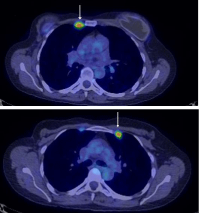
    Figure 1: Axial FDG PET/CT: FDG uptake in bilateral IM (arrows) nodes and
axillary nodes (not shown) highly suspicious for breast cancer metastases.
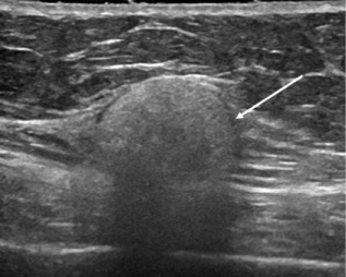
    Figure 2: Right Axially Ultrasound: US of the right axilla demonstrate a
typical snowstorm appearance of silicone within an axillary node (arrow).
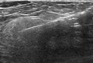
    Figure 3: US-Guided Biopsy: Ultrasound guided biopsy of aright axillary
lymph node was performed which revealed silicone granuloma.
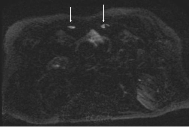
    Figure 4: Axial Silicone Selective MR pulse sequence: Subtle high signal
intensity in IM nodes suggestive of silicone (arrows).
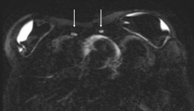
    Figure 5: Axial Silicone Selective MR pulse sequence: Dual lumen
implants are intact. Subtle high signal in IM nodes (arrows) suggestive of
silicone, partially obscured by pulsation artifact.
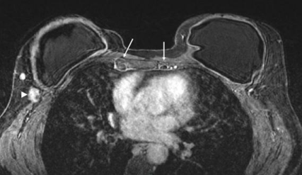
    Figure 6: Axial T1 post contrast MRI: Enlarged low signal intensity IM
nodes (arrows); low intensity on post-gad due to silicone. Note an enlarged,
enhancing right axillary node (arrowhead).
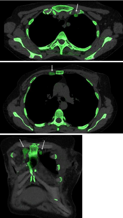
    Figure 7: Axial and Coronal Dual-Energy CT: Multiple enlarged IM lymph
nodes (arrows) containing silicone (green color mapping) corresponding to
the PET/CT.
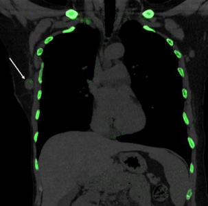
    Figure 8: Coronal DECT: Small amount of silicone noted in an enlarged right
axillary node (arrow).
Discussion
The prevalence of silicone breast implant rupture is quite high, estimated to be 6% per year in a cumulative fashion for the first five years following implant surgery [1] and estimated to be as high as 77% over the lifetime of the implant [2]. More recent studies evaluating modern silicone implants suggest the rupture rate may be reasonably lower, around 15% during the first three to ten years. Nonetheless, implant rupture remains a not uncommon problem.
Complications from implant rupture can include pain, contractures and cosmetic asymmetry. However, most patients with ruptured silicone implants are asymptomatic; the silent rupture rate is approximately 55% [3]. While local complications are troubling for the patient, we report a case of diagnostic uncertainty which was troubling for the radiologist and clinical team.
Our patient’s history of a childhood Wilm’s tumor treated with radiation extending from the right shoulder to the right hip put her at a significantly elevated risk for developing breast cancer, reported as high as 30% by the age of 50 years [4]. This elevated risk has led to the accepted practice of screening women who received chest irradiation =20 Gy as children or young adults with annual mammography and breast MR starting at the age of 25 years or 8 years after radiation treatment. When our patient presented to her clinical team with new nonspecific symptoms (fatigue, headaches and shooting breast pain), an imaging workup appropriately began. PET/CT exam showed hypermetabolic lymph nodes in her right axilla, right subpectoral region, and in her bilateral internal mammary lymph node chains, suspicious for breast cancer recurrence.
It has been well documented that extracapsular silicone can migrate to regional lymph nodes; this is felt to occur either by direct migration or by macrophagic phagocytosis of silicone microdroplets which subsequently travel to regional lymph nodes [5]. While it has been previously reported that silicone granulomatous lymph nodes can lead to false positive PET/CT findings [6], additional confirmatory test were warranted in this case to exclude other ominous etiologies.
An additional unique and confounding feature of this case was the contralateral internal mammary chain involvement. While rare, a few prior case reports have demonstrated contralateral IM node involvement with silicone granulomas [5,7,8]. Furthermore, Vendrell-Town investigated the physiologic lymphatic drainage pathway in 250 healthy women using radioactive isotopes and found a rate of 0.8% (2/250) demonstrating contralateral IM nodal drainage [9].
Fortunately, the patients axially nodes were easily amenable to ultrasound guided fine needle aspiration which revealed silicone granulomas (subsequently confirmed with core needle biopsy). The suspicious subpectoral and bilateral internal mammary lymph nodes likely represented the same process, however additional confirmatory imaging was sought and the patient subsequently had breast MR imaging preformed.
Silicone sensitive MR sequences are highly accurate at characterizing extracapsular silicone in breast parenchyma, however there is pulsation artifact which can obscure identification of silicone within level I through III axillary lymph nodes or internal mammary nodes. The supraclavicular nodes may not be included on the MR Field of view. However not all patient can tolerate MR imaging (incompatible pacemaker, claustrophobia, finically prohibited, etc.) and DECT may offer a more suitable alternative.
DECT uses CT numbers (measured in Hounsfield units) which depend on X-ray attenuation which depends on physical density (g/ cm3) and atomic number (Z). Silicone contains the atomic element silicon which has an atomic number of 14, while soft tissue is predominantly made up of lighter elements including hydrogen (atomic number 1) and oxygen (atomic number 8). The differences in atomic number result in different slopes in a plot of low energy CT number versus high energy CT number which can be used to characterize differences in tissue composition. Our patients DECT scan clearly demonstrated silicon within all of the suspicious lymph nodes identified on PET/CT imaging. Confidently making the diagnosis of silicone granulomatous lymph nodes on imaging alone could have spared our patient two biopsy procedures.
MRI remains the gold standard for detecting extracapsular silicone, however it may be more difficult to identify within regional lymph nodes. DECT demonstrates potentially equal diagnostic accuracy in detecting extracapsular spread of silicone particularly in level II and III axillary nodes and internal mammary nodes. In our patient, we were able to confidently detect silicone in the regional lymph nodes on DECT, accounting for the abnormal uptake on the PET/CT imaging.
References
- Marotta JS, Goldberg EP, Habal MB, Amery DP, Martin PJ, Urbaniak DJ, et al. Silicone gel breast implant failure: Evaluation of properties of shells and gels for explanted prostheses and meta-analysis of literature rupture data. Ann Plast Surg. 2002; 49: 227–247.
- Leduey A, Mazouni C, Leymarie N, Heba Alkhashnam, Benjamin Sarfati, Jean-Rémi Garbay, et al. Comparison of the explantation rate of poly implant prothèse, Allergan, and Pérouse silicone breast implants within the first four years after reconstructive surgery before the poly implant prothèse alert by the French Regulatory Authority. Int J Breast Cancer. 2015; 2015: 519497.
- Brown SL, Middleton MS, Berg WA, Soo MS, Pennello G. Prevalence of rupture of silicone gel breast implants revealed on MR imaging in a population of women in Birmingham, Alabama. Am J Roentgenol. 2000; 175: 1057-1064.
- Moskowitz CS, Chou JF, Wolden SL, Bernstein JL, Malhotra J, Novetsky Friedman D, et al. Breast cancer after chest radiation therapy for childhood cancer. J Clinical Oncol. 2014; 32: 2217–2223.
- Gil T, Mettanes I, Aman B, Taran A, Shoshani O, Best LA, et al. Contralateral internal Mammary Silicone Lymphadenopathy Imitates Breast Cancer Metastasis. Ann Plastic Surgery. 2009; 63: 39-41.
- Ataergin S, Arslan N, Ozet A, Ozguven MA. Abnormal 18F-FDG Uptake Detected with Positron Emission Tomography in a Patient with Breast Cancer: A Case of Sarcoidosis and Review of the Literature. Case Reports in Medicine. 2009; 785047.
- Kao CC, Rand RP, Holt CA, Pierce RH, Timmons JH, Wood DE. Internal mammary silicone lymphadenopathy mimicking recurrent breast cancer. Plast Reconstr Surg. 1997; 99: 225–229.
- Glazebrook KN, Leng S, Jacobson SR, McCollough CM. Dual-energy CT for evaluation of intra- and extracapsular silicone implant rupture. Case Reports in Radiol. 2016, Article ID 6323709.
- Vendrell-Torne E, Setoain-Quinquer J, Domenech-Torne FM. Study of normal mammary lymphatic drainage using radioactive isotopes. J Nucl Med. 1972; 13: 801-805.