
Case Report
Austin J Clin Case Rep. 2017; 4(3): 1123.
Painful Congenital Coracoclavicular Joint, Treated with Arthroscopic Resection: A Case Report
Kulkamthorn N and Phonphok P*
Division of Sports Medicine, Department of Orthopedics, Phramongkutklao Hospital and College of Medicine, Thailand
*Corresponding author: Phonphok P, Division of Sports Medicine, Department of Orthopedics, Phramongkutklao Hospital and College of Medicine, Bangkok10400, Thailand
Received: June 26, 2017; Accepted: July 24, 2017; Published: August 04, 2017
Abstract
A case of painful right shoulder in a 39 year old Thai female. She had a painful range of motion due to forward flexion and adduction with no any limit active range of motion for 5 months. The diagnosis was confirmed with incisional biopsy for tissue pathological report. 9-months after treated with arthroscopic coracoclavicular joint resection, she regained a pain-free range of motion.
Introduction
The coracoclavicular joint is an uncommon anatomical variant with a diarthrotic synovial joint between the conoid tubercle of the clavicle and the superior surface of the coracoid process [1]. From the previous studies, the prevalence varies between 0.04–30% and much more higher in Asia than in Europe and Africa [2-5]. By the way the coracoclavicular joint does not usually produce any symptom [6-7].
Case Presentation
A 39-year old Thai female presented with her severe right shoulder pain for 5 months, without any traumatic history before. The right shoulder examination revealed full active and passive range of motion. The pain was aggravated when she forward flexion and adduction her shoulder. She can’t able to do any heavy weight lifting due to her painful shoulder. The radiographic examination revealed the presence of an accessory joint between the coracoid and the conoid tubercle (Figure 1). The computerized tomography (Figure 2) and the Magnetic resonance imaging study reveal the presence of a pseudarthrosis with degenerative diarthrosis between the coracoid and the distal end of the clavicle (Figure 3).
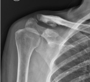
    Figure 1:  Radiographic showing accessory joint between the coracoid and
the conoid tubercle.
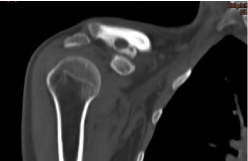
    Figure 2:  CT scan at the level of the coracoclavicular joint confirms the
degenerative changes in the joint surfaces.
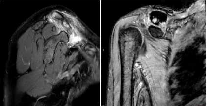
    Figure 3:  MRI reveal the presence of a pseudarthrosis between the coracoid
and the distal end of the clavicle.
The incisional biopsy was done through the pathological lesion and mature hyaline cartilage was reported from the study. The local 2% xylocaine injection test was performed at the coracoclavicular joint to confirmed the source of her pain. Dramatic pain relief at her shoulder was noted after xylocaine injection was done. However, the pain was relieved for just only a few hours and the pain was still the same as before the procedure.
Treatment
After we decided to treat the patient initially by non-operative treatment. Pain management was done by oral anti-inflammatory medications (NSAIDs) and local corticosteroid injection. We also advise the patient to restrict her weight lifting activity and avoid all activities that stress the AC joint, such as pushing and pulling. 6 months after the non-operative treatment the pain was improved but still disturbs her daily life activity. Arthroscopic resection of the coracoclavicular joint was performed in order to improve her life quality by restore her shoulder motion. The procedure was performed in standard beach chair position with a cushion under the scapula of the affected shoulder by under controlled hypotension and a combination of regional and general anesthesia for better visualization and post-operative recovery.
A standard posterior portal was created and the arthroscope was inserted into the glenohumeral joint. An anteroinferior portal was created with an outside-in technique through the rotator cuff interval. The anterior capsular structures are resected until the coracoid was well visualized. The anterosuperiorportal is then identified and created with a spinal needle at a point just anterior to the anterior margin of the acromion. Then the coracoid and also the coracoclavicular joint can be identified directly after the scope was transferred to the anterosuperior portal (Figure 4). The coracoclavicular joint was resected through the anteroinferior portal by arthroscopic burr (Figure 5). The resection begins laterally and proceeds medially. The margin and adequate of coracoclavicular joint resection was checked by using a fluoroscope (Figure 6) and reference with the inferior margin of the clavicle (Figure 7).
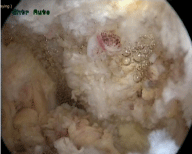
Figure 4: The coracoclavicular joint viewing from the anterosuperior portal.
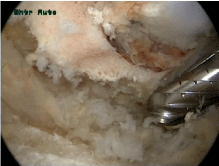
Figure 5: Arthroscopic resection of the joint through the anteroinferior portal.
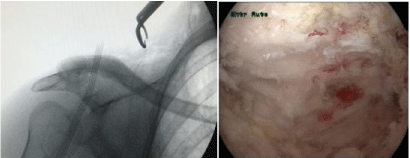
    Figure 6 and 7:  The resection margin was checked by using a fluoroscope
and reference with the inferior margin of the clavicle.
After the operation her arm was placed in a sling for 2 weeks after that she was allowed to begin her isometric shoulder strengthening and active shoulder range of motion exercised. The range of motion exercise up to flexion and abduction of 45 degree in the first 2 weeks and up to 90 degree in next 3 weeks. At 8 weeks we used the resistance band to performed the periscapular muscle strengthening exercises. At nine months postoperatively, she regained her full active range of motion with no painful arc of motion at her shoulder at all. No evidence of recurrence or inadequate coracoclavicular joint resection was seen from the postoperative radiographic study anymore (Figure 8).

    Figure 8:  Postoperative radiographic showing no evidence of inadequate or
recurrence coracoclavicular joint formation.
The acromioclavicular joint was clinically stable with full active range of motion of her shoulder at 18 months postoperatively. There was no prominence, subluxation, or dislocation of the acromioclavicular joint (Figure 9).
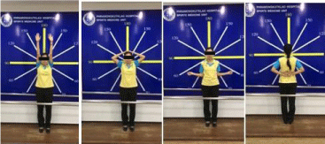
    Figure 9:  At18 months postoperatively. The acromioclavicular joint was
stable with full active shoulder motion.
Discussion
A coracoclavicular joint is a rare anatomical entity that is more common in Asians than in other races. From the previous study, the prevalence ranges between 0.7–10% in osteological studies, 1.7- 30% in cadaveric dissections and 0.04-3.0% in radiographic studies. But normally this anomalous joint dose not usually produces a painful arch of motion [6-7]. Conservative treatment is still the first-line of treatment. However if conservative treatment was failed, coracoclavicular joint excision is a good surgical option. The excision can perform by both open and arthroscopic technique [8]. In our case, we resected the joint by arthroscopic technique with a good postoperative clinical and radiographic results, no painful arch of motion in just 10 months postoperatively. We recommend arthroscopic excision technique in the patient who need less postoperative recovery time.
References
- Gradoyevitch B. Coracoclavicular joint. J Bone Joint Surg. 1937; 21: 918- 920.
- Cockshott WP. The geography of coracoclavicular joints. Skeletal Radiol. 1992; 21: 225-227.
- Nehma A, Tricoire JL, Giordano G, Rouge D, Chiron P, Puget J, et al. Coracoclavicular joints. Reflections upon incidence, pathophysiology and etiology of the different forms. Surg Radiol Anat. 2004; 26: 33-38.
- Gumina S, Salvatore M, De Santis R, Orsina L, Postacchini F. Coracoclavicular joint: osteologic study of 1020 human clavicles. J Anat. 2002; 201: 513-519.
- Nalla S, Asvat R. Incidence of the coracoclavicular joint in South African populations. J Anat. 1995; 186: 645-649.
- Possati A. Uncasodiarticolazionecoraco-clavicolareosservatoradiograficame nte. Chir d Org di movimento. 1926; 10: 533-536.
- Timpano M. Aspettiradiografici dell’ articolazionecoracoclavicolare. Ann di Radiol e Fis Med. 1934; 491-507.
- Singh VK, Singh PK, Balakrishnan SK. Bilateral coracoclavicular joints as a rare cause of bilateral thoracic outlet syndrome and shoulder pain treated successfully by conservative means. Singapore Med J. 2009; 50: 214-217.