
Case Report
Austin J Clin Case Rep. 2019; 6(5): 1159.
Atypical Fibrous Histiocytoma: A Case Report
Singer R1*, Kiziltaç K1, Etikan P2 and Erdem ZB3
¹Okmeydani Training and Research Hospital, Department of Dermatology, Istanbul, Turkey
²Dr. Akçiçek State Hospital, Department of Dermatology, Girne, Cyprus, Turkey
³Okmeydani Training and Research Hospital, Department of Pathology, Istanbul, Turkey
*Corresponding author: Ralfi Singer, Department of Dermatology, Okmeydani Training and Research Hospital, Darülaceze Cad. No: 25, 34384, Istanbul, Turkey
Received: November 25, 2019; Accepted: December 26, 2019; Published: December 31, 2019
Abstract
Atypical fibrous histiocytoma is a rare variant of dermatofibroma, which may present diagnostic problems because of histopathological features, which are difficult to distinguish from a malignant tumor. The neoplasm has a tendency to recur locally and a capacity to metastasize, although very rarely. Here, we report a 46-year-old man who presented with a firm dermal nodule on the right knee. The diagnosis was made on the basis of clinical, histopathological and immunohistochemical findings. We discuss the clinical and histopathological features as well as the differentiating features of atypical fibrous histiocytoma from other cutaneous spindle cell tumors such as dermatofibrosarcoma protuberans, atypical fibroxanthoma and pleomorphic dermal sarcoma.
Keywords: Atypical fibrous histiocytoma; Dermatofibroma; Spindle cell tumors
Introduction
Fibrous histiocytoma (dermatofibroma, sclerosing hemangioma, nodular subepidermal fibrosis) (FH) is a common benign cutaneous neoplasm of the skin [1]. Many histological variants of FH such as aneurysmal, atypical, cellular, epithelioid, keloidal, atrophic, hemosiderotic, lipidized, palisading, clear cell and FH with osteoclastlike giant cells subtypes have been identified over the years [1,2]. Atypical fibrous histiocytoma (AFH) is an uncommon variant of FH; the tumor is a diagnostic challenge because of histopathological features which are difficult to differentiate from a malignant tumor [1,3]. Because of this problem, it needs wider recognition in order to preclude unsuitable management. Herein, we present a case of AFH located to the lower extremity.
Case Presentation
A 46-year-old man presented to our outpatient clinic with a 5-month history of a painful, slowly growing lesion located on the right knee (Figure 1). The medical history was not remarkable. On physical examination, a 1.5cm violaceous nodular lesion with a firm consistency was noted. The lesion was totally excised and histopathological examination demonstrated spindle cells with atypical nuclei arranged in a storiform pattern in the dermis. Pleomorphic giant cells with large bizarre nuclei were noted as well (Figure 2). Immunohistochemical staining demonstrated no staining for cytokeratin, CD31, CD34, HMB 45, S-100, desmin and smooth muscle actin. Vimentin staining was not performed and Ki 67 staining showed 20% positivity. AFH was diagnosed on the basis of these findings. Since the patient was lost for follow-up, we could not perform radiologic screening to search for possible metastasis. An informed consent has been obtained from the patient to share the data in a scientific journal.
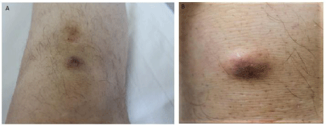
Figure 1A and 1B: Violaceous nodular lesion located to the right knee.
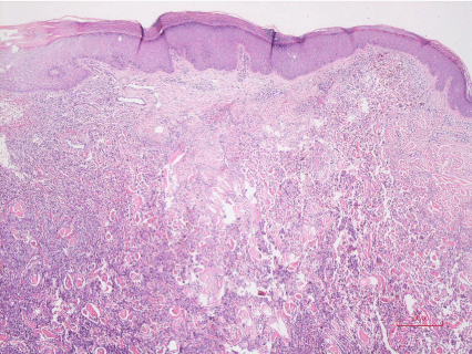
    Figure 2A: Hyperplastic epidermis and atypical spindle cell infiltration
comprising superficial and deep dermis (HE; x40).
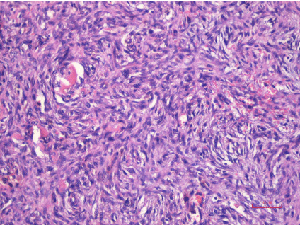
    Figure 2B: Spindle cells and oval-round cells with atypical nuclei and
prominent nucleoli arranged in a storiform pattern and increased mitotic
activity. Neoplastic tissue forms clefts in the collagen (HE; x200).
Discussion
FH is the most frequent mesenchymal neoplasm of the skin. AFH is a rare variant of cutaneous FH which presents as a solitary firm cutaneous nodule in wide age range (5 to 79 years) most commonly located to the lower and upper extremities [2,4]. The synonyms are pseudosarcomatous fibrous histiocytoma or dermatofibroma with monster cells [2]. Although it has a low malignant potential, it shows a higher persistence rate after limited removal in comparison to dermatofibroma. Metastases are rare [4,5]. Nevertheless, AFH has a higher tendency to recur locally akin to the aneurysmal and cellular variants of FH [4].
Histologically, AFH shows proliferation of dermal spindle cells consisting mainly of fibroblast-like spindle cells and atypical histiocytic cells in a background of classic fibrous histiocytoma [3,4,6]. Typical features are pleomorphic, round, spindle and/or polyhedral cells with large hyperchromatic, irregular nuclei, bizarre multinucleated cells (monster cells) and xanthomatous cells with large prominent nuclei [4,6]. Usually, a grenz zone is present. Superficial part of the subcutis may be involved in one third of the cases. Immunohistochemical staining demonstrates diffuse positivity for vimentin; CD34, alpha smooth muscle actin, and desmin are sometimes focally positive. CD68 and factor XIIIa positivity are variable [2].
AFH should be distinguished from other spindle cell tumours such as dermatofibrosarcoma protuberans (DFSP), atypical fibroxanthoma (AFX), pleomorphic dermal sarcoma (PDS) and dermal leiomyosarcoma [3,7]. DFSP demonstrates uniform spindle cells having spindle-shaped nuclei, which demonstrate a storiform pattern. The tumor extends from the dermis to the subcutaneous tissue with an indistinct border; immunohistochemical analysis reveals diffuse CD34 positivity [3]. AFX usually presents on the head and neck of elderly patients. Although AFX lacks the typical characteristics of FH, it has similar features to those of AFH in showing proliferation of spindle cells, multinucleated giant cells and bizarre cells. However, AFX is characterized by solar elastosis, no grenz zone is seen and the tumor does not extend into subcutis [3,4]. The immunohistochemical features of AFH and AFX are similar [7]; both the spindle-shaped cells and the histiocyte-like cells show positive reaction for vimentin. The spindle-shaped cells react positively for muscle specific actin, whereas the large histiocyte-like cells are positive for CD68 and CD163 [8]. MiB1 helps to distinguish these two entities since AFX shows a very high rate of proliferating atypical cells [9]. Although PDS demonstrates similar histopathological and immunohistochemical characteristics to those of AFX, it shows deeper infiltration, necrosis, vascular or perineural invasion. PDS and AFX are diagnosed by excluding other spindle cell and pleomorphic neoplasms because of their overlapping histopathological features and no specific immunohistochemical or molecular markers can be used for the differential diagnosis. Nevertheless, immunohistochemistry is necessary for the diagnosis of PDS or AFX, because the absence of cytokeratin, desmin, S100 and CD34 expression is a diagnostic criterion [10]. Dermal leiomyosarcomas are superficial malignant smooth muscle neoplasms which are usually poorly circumscribed and are made of spindle cells arranged in fascicles that infiltrate collagen bundles. Immunophenotypic studies usually show smooth muscle actin and desmin positivity [7,8]. DFSP and AFX have intermediate malignancy whereas PDS and dermal leiomyosarcomas are more aggressive neoplasms [3].
Conclusion
It is important to include this rare variant of fibrous histiocytoma in the differential diagnosis of dermal mesenchymal neoplasms and it should be totally excised with clear margins because of the risk of local recurrence and the rare occurrence of metastasis.
References
- Ishitsuka Y, Ohara K, Otsuka F. Atypical fibrous histiocytoma of the skin with necrobiotic granuloma-like features. Acta Dermato Venereol. 2011; 91: 482- 483.
- Szablewski V, Laurent-Roussel S, Rethers L, Rommel A, Van Eeckhout P, Camboni A, et al. Atypical fibrous histiocytoma of the skin with CD30 and p80/ALK1 positivity and ALK gene rearrangement. Journal of Cutaneous Pathology. 2014; 41: 715-719.
- Tsunoda K, Takahashi K, Maeda F, Oikawa H, Akasaka T. A case of atypical fibrous histiocytoma with positivity for CD163 and CD44. Acta Derm Venereol. 2013; 93: 737-738.
- Kaddu S, McMenamin ME, Fletcher CDM. Atypical fibrous histiocytoma of the skin: clinicopathologic analysis of 59 cases with evidence of infrequent metastasis. Am J Surg Pathol. 2002; 26: 35-46.
- Guillou L, Gebhard S, Salmeron M, Coindre JM. Metastasizing fibrous histiocytoma of the skin: a clinicopathologic and immunohisto-chemical analysis of three cases. Mod Pathol. 2000; 13: 654-660.
- Huan Y, Vapnek J, Unger PD. Atypical fibrous histiocytoma of the scrotum. Ann Diagn Pathol. 2003; 7: 370-373.
- Ben Abdelkrim S, Belajouza C, Jomaa W, Beizig N, Ben Said Z, Mokni M, et al. Atypical Cutaneous Fibrous Histiocytoma: An Unusual and Misleading Variant of Fibrous Histiocytoma. Case Rep Pathol. 2011; 2011: 612416.
- Calonje E. Soft tissue tumours and.tumours-like conditions. In: Rook’s Textbook of Dermatology. Burns T, Breatnach S, Cox N, Griffiths C, editors. 8th ed. Oxford: Wiley-Blackwell. 2010: 56.1-56.62.
- Wilk M, Zelger BG, Nilles M, Zelger B. The value of immunohistochemistry in atypical cutaneous fibrous histiocytoma. Am J Dermatopathol. 2004; 26: 367-371.
- Miller K, Goodlad JR, Brenn T. Pleomorphic dermal sarcoma: adverse histologic features predict aggressive behavior and allow distinction from atypical fibroxanthoma. Am J Surg Pathol. 2012; 36: 1317-1326.