
Research Article
Austin J Clin Neurol 2016; 3(1): 1085.
Evaluation of Lung, Cardiac and Brain Pathologies of Death Cases Caused by Carbon Monoxide Poisoning
Pehlivan S¹*, Alacam H², Samdanci E³, Kara D¹, Turkkan D¹, Altuntas A4, Gurler M² and Karapirli M5**
¹Department of Pathology- Morgue, Ankara Branch of the Council of Forensic Medicine, Turkey
²Department of Medical Biochemistry, Hacettepe University, Turkey
³Department of Pathology, Inonu University, Turkey
4Department of Chemistry, Ankara Branch of the Council of Forensic Medicine, Turkey
5Department of Forensic Medicine, Harran University, Turkey
*Corresponding author: Sultan Pehlivan, Department of Pathology - Morgue, Ankara Branch of the Council of Forensic Medicine, Ankara ŞefkatMah. Felek Cad. No: 45 Keçiören/Ankara 06300, Turkey
**Senior author: Mustafa Karapirli, Department of Forensic Medicine, School of Medicine, Harran University, Sanliurfa, Turkey
Received: April 14, 2016; Accepted: May 30, 2016; Published: June 02, 2016
Abstract
Background: Carbon monoxide (CO) poisoning is a big problem and seen in the whole world. And, the pathological changes caused by CO poisoning is still investigated.
Aim: The aim of this study is to investigate the relationship between pathological changes and carboxyhemoglobin (COHb) levels.
Method: 127 death cases, due to CO poisoning, are included in the study. Lung tissue samples are examined histopathologically and evaluated in terms of edema, hemorrhage, congestion, pneumonia, bronchitis, emphysematous changes, foreign body aspiration and pleural pathology. Hemorrhage, edema and other specific findings in the brain are evaluated. For examination of cardiac tissue, secondary findings in the sectional myocardial ischemia and stenosis in the coronary artery sections are evaluated. And, the correlation between the pathological changes and COHb levels were investigated.
Results: Edema (80%), hemorrhage (27%), congestion (13%), emphysematous changes (42%), pneumonia (0.03%) and bronchitis (0.05%) were detected in the lungs. Cardiac pathologies such as coronary stenosis (11%), cardiac ischemia (0.007%) and fibrosis (0.08%) and brain lesions (0.03%) were located in the cases. However, no correlation between changes in cardiac and brain pathology with COHb levels were detected. A negative correlation was found between COHb with pneumonia and bronchitis.
Conclusions: As a result, besides damage in the central nervous system and heart, severe damage also occurs in the lungs and various clinical conditions such as edema, congestion and pneumonia can be seen. All these histopathologic findings may be helpful to consider planning of acute and long term treatment of CO poisoning.
Keywords: CO poisoning; Carboxyhemoglobin; Pulmonary edema; Pulmonary congestion
Introduction
Carbon monoxide (CO) occurs from incomplete combustion of organic substances and it is a toxic gas with the features of colorless, odorless and non-irritating. CO is also formed during the destruction of the hem group in the human body and through the binding of hemoglobin (Hb), carboxyhemoglobin (COHb) arises. The COHb level in healthy individuals lies between 1-3%. But the COHb levels in the blood can reach up to 10-15% in persons that smoke a lot [1-4].
CO poisoning makes up nearly half of all deadly poisonings in the world [5,6]. Non-intentional CO poisoning takes place in different regions and different seasons in the world [7]. CO poisoning in Turkey is the most common cause of morbidity and mortality in the winter months. Also, considering all age groups, CO poisoning is the first reason of fatal poisonings in Turkey [8-11].
Pathological findings and clinical manifestations after CO poisoning depend on the CO exposure conditions. CO exposure can differ in amount and time. Short or long term exposure to high concentrations of gas or long term exposure to low concentrations of gas can cause CO poisoning [12].
CO shows its effect via hypoxia, cellular toxicity and free radicalmediated cellular injury. By binding itself to hemoglobin, CO slides the oxygen dissociation curve to the left which leads to less oxygen separating from the hemoglobin resulting in cellular hypoxia. Cellular toxicity arises through the connection of CO with different proteins and enzymes inside the cell. Especially, cytochrome c oxidase connected CO hinders the cells to use oxygen by the mitochondria. When electrons don’t reach the oxygen, they are separated from the carrier and cause free radical-mediated cellular injury inside the cell. An increase in lipid peroxidation resulting from the interaction of unsaturated fatty acids with oxygen free radicals after CO poisoning in rats [13,14].
During CO poisoning, depending on the impact level of hypoxia, different findings can arise. If the central nervous system is affected, headache, syncope, loss of consciousness varying from disorientation to coma, seizures, ataxia, behavioral disorders, dizziness, balance disorders and general weakness are observed. If the cardiovascular system is affected, palpitations, feeling of tightness in chest, cardiac arrhythmia and ischemic changes are especially seen in individuals with coronary artery diseases. Cardiorespiratory arrest can arise due to brainstem changes or severe cardiac hypoxia [15,16]. Besides the changes in the nervous and cardiac system, different levels of lung pathologies can also be observed. Pulmonary changes seen in CO poisoning are connected to prolonged hypoxia. These changes in the lungs are affected by the permeability of capillaries and cause edema [17,18]. Adult respiratory distress syndrome is rarely seen after CO poisoning [19]. Although all of these histopathological information’s related with CO poisoning in cardiac, lung and brain tissues, there are still some deficiencies in this area. Therefore, the aim of this study is to investigate the relationship between pathological changes in especially postmortem lung, heart and brain tissues and their COHb levels.
Materials and Methods
This is a retrospective study. Work permit was received by the Commission for Education and Scientific Research of the Forensic Medicine Institution (Date: March 12, 2013, number: B.03.1.ATK.0.01.00.08/157)
Cases
One hundred and twenty seven (127) death cases that resulted from CO poisoning, between the years of 2007 and 2012, were included in the study. Seventy (70) of those are men and 57 women. Demographic characteristics of the cases are shown in Table 1. Just, CO poisoning cases included in the study. The autopsies of the cases were conducted at the Institute of Forensic Medicine in the Morgue Department of the Ankara Group Presidency.
![]()
    Age
    Sex
    Total


    Women
    Men


    0-10
    6
    3
    9


    11-20
    5
    5
    10


    21-30
    6
    10
    16


    31-40
    3
    6
    9


    41-50
    6
    9
    15


    51-60
    8
    10
    18


    61-70
    9
    10
    19


    71-80
    8
    11
    19


    81-90
    6
    6
    12


    Total
    57
    70
    127
Table 1: The table shows the age and sex of the cases.
Histopathological examination
Lung, heart and brain tissue samples from the cases were examined histopathologically. Lung tissue samples were examined histopathologically and evaluated in terms of edema, hemorrhage, congestion, pneumonia, bronchitis, emphysematous changes, foreign body aspiration and pleural pathology. Hemorrhage, edema and other specific findings in the brain tissue were evaluated. Coronary stenosis, ischemia, fibrosis and other specific findings were noted in the heart tissue. Table 2 displays which pathologies were scored in what way.
![]()
    Pathological State
    Scores


    0
    1
    2
    3


    Lung edema
    Absent
    Focal edema
    Alveolar edema


    Hemorrhage in the lungs
    Absent
    Focal hemorrhage
    Alveolar hemorrhage


    Lung congestion
    Absent
    Existent


    Pneumonia
    Absent
    Existent


    Bronchitis
    Absent
    Bronchitis
    Bronchopneumonia


    Emphysematous changes
    Absent
    Existent


    Foreign body aspiration
    Absent
    Existent


    Brain lesion
    Absent
    Existent


    Coronary stenosis
    Absent
    30-33% narrowing
    34-66%
    67-100%


    Cardiac ischemia
    Absent
    Perivascular ischemia
    Interstitial ischemia
    Fibrosis/scarring


    Cardiac hypertrophy
    Absent
    Existent
Table 2: The table shows how lung, cardiac and brain pathologies of the autopsy cases are scored.
Biochemical parameters
Besides the result of the tissue examination also postmortem carboxy hemoglobin (HbCO, % saturation) levels of the cases were noted. According to the COHb levels the cases were divided into three groups:
Group 1: COHb levels were between 0-29.9 %
Group 2: 30-59.9 %
Group 3: 60-90 %.
Statistical analyses
The data of the study didn’t show normal distribution. The correlation analyses were carried out with Spearman’s correlation analysis. Binary comparisons were carried out with the Mann- Whitney U test. p≤ 0.05 was considered significant.
Results
Demoghraphic data
The average age of all cases, men and women were respectively 49.2, 49.47 and 48.87. The age of men was between 1 and 85 and the age of women between 1 and 88.
Because some autopsies were conducted outside the center of the study and their files were kept there, we couldn’t reach the cause of death. The causes of death of sixty five (65) cases are presented in Figure 1. The most common reason is stove poisoning.
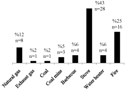
    Figure 1: Causes and percentage of poisoning. The biggest reason is stove
poisoning.
Examination of lung, cardiac and brain tissues
The all pathologies seen in lung, cardiac and brain tissues were showed in Table 3. The most common lung pathology was pulmonary edema among male and female. The second common seen was emphysematous changes. While coronary stenosis was the most common cardiac pathology among female, fibrosis was the most among men (Table 3).
![]()
    Tissue
    Histopathologic parameters
    Male(%)
    Female (%)
    Total (%)


    Lung
    Edema
    76
    84
    80


    Hemorrhage
    30
    23
    27


    Congestion
    11
    16
    13


    Pneumonia
    0.01
    0.05
    0.03


    Bronchitis
    0.04
    0.05
    0.05


    Emphysematous changes
    43
    40
    42


    Foreign body aspiration
    0.09
    0.04
    0.06


    Pleural pathology
    0.01
    0
    0.007


    Cardiac
    Coronary stenosis
    0.04
    19
    11


    Ischemia
    0.01
    0
    0.007


    Fibrosis
    10
    0.05
    0.08


    Other specific findings
    0.04
    0.02
    0.03


    Brain pathologies(subarachnoid hemorrhage, superficial punctate    bleeding, edema)
    0.03
    0.04
    0.03
Table 3: Pathologies in the lung, cardiac and brain tissues.
Pleural changes in lung pathology was only detected in a case of a 14 year old, in form of acute fibrinous pleuritis.
Examining the relationship between age and COHb levels didn’t reveal a significant relationship (p=0.173 and r=-0.122).
While examining the relationship between COHb and lung parameters hasn’t shown a significant relationship in COHb and lung edema (p=0.434, r=0.070), hemorrhage (p=0.307, r=-0.091), congestion (p=0.227, r=-0.108), emphysematous changes (p=0.944, r=0.006) and foreign body aspiration (p=0.661, r=-0.039), a negative correlation was determined between COHb and pneumonia (p=0.027, r=-0.196) and bronchitis (p=0.006, r=-242) (Figure 2, 6, 7).
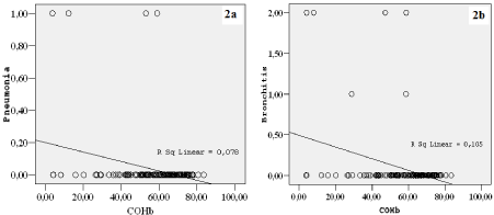
Figure 2: The graph shows the relationship between COHb and pneumonia. Negative correlation draws attention on both graphs.
No significant relationship was found between COHb and brain pathologies (p=0.179, r=-0.120).
Also no significant relationship was found between COHb and cardiac pathologies (coronary stenosis, cardiac ischemia and cardiac hypertrophy) (coronary stenosis: p=0.941, r=-0.007; cardiac ischemia: p=0.641, r=0.042; cardiac hypertrophy: p=0.414, r=-0.073)
There was a positive correlation between age and coronary stenosis (p<0.001, r=0.308) (Figure 3).
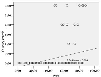
    Figure 3: The graph shows the relationship between age and coronary
stenosis.
When we look at the relationship between the lung parameters, there was a negative correlation between the lung edema and emphysematous changes (p=0.018, r=-0.21). Similar to that, there was a negative correlation between congestion and emphysematous changes (p<0.001, r=0.333) (Figure 4, 6, 7).
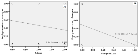
Figure 4: The graph shows the relationship between congestion and emphysematous changes.
There was a positive correlation between bronchitis and brain pathologies (p<0.001, r=0.391) and coronary stenosis (p=0.001, r=0.296) (Figure 5).
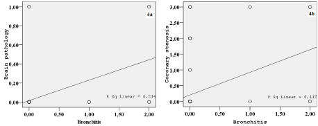
Figure 5: The graph shows the positive correlation between bronchitis and brain pathologies and coronary stenosis.
There was a matter of positive correlation between foreign body aspiration and emphysematous changes (p=0.006, r=0.241) (Figure 6, 7).
In addition to the results above, as expected, there was a positive correlation between coronary stenosis and cardiac ischemia (p<0.001, r=0.522) and a positive relationship between cardiac ischemia and cardiac hypertrophy (p<0.001, r=0.429).
Discussion
Most important sources of CO gas are wrongly installed heaters, motor vehicles, devices using carbon fuels and house fires. In Turkey; however, the most common reason for CO poisoning is the stove which is closely followed by fires caused by water heaters [7,20]. If the heating device is poorly or wrongly installed and the ventilation of the environment is inadequate, it can very quickly lead to death. Also CO poisoning arising from a fire can lead to sudden deaths [21]. The most common cause of death in our study was stove poisoning followed by fires.
The most important factor determining the intoxication level in CO poisoning is the inhaled CO amount and exposure time [22]. Healthy individuals can live 1 minute with a 40% COHb amount and 1 week with a 20% COHb amount. The COHb level can rise up to 60% and higher in a subject exposed to smoke of an internal combustion engine which is lethal [23]. The COHb levels of only 11 cases in our study were below 30%. The rest of the 116 cases had COHb levels over 30%. This shows that the COHb level in cases where death occurs is rather high.
CO binds to hemoglobin with a high affinity and creates COHb. CO also disrupts the function of the electron transport chain and causes that the electrons are set free. Thus, the occurring free oxygen radicals are causing damage to the organic compounds within the cell. The fact that CO binds to myoglobin in the skeletal muscle with three times higher affinity contributes to extra damage on a cellular level. It is thought that brain lipid peroxidation plays an important role in neurological manifestations during CO intoxication [13,24].
Due to their high metabolic rate the brain and heart are most vulnerable to CO-induced hypoxia [22]. Therefore, the death is often resulted from cardiac. When the cardiovascular system is affected, changes such as palpitations, feeling of tightness in the chest, severe hypotension, fatal arrhythmias, electrocardiographic changes, pulmonary edema and especially ischemic cardiac changes in individuals with coronary artery disease are seen [6,15,20,25-27]. In a study carried by Korb et al., it is reported that a myocardial ischemia is seen after an at least 24 hour latent period [28]. In our study we have found an ischemia diagnosis after 3-7 days in a case that has resulted in death after a 7-day hospital treatment. If the central nervous system is affected; headache, syncope, fainting loss of consciousness ranging from disorientation to coma, seizures, ataxia, behavioral disorders, dizziness, balance disorders, general weakness and neuropsychiatric symptoms are observed. In a study from Akdemir et al., 12 cases of 55 CO poisoning has shown increased levels of troponin because of myocardial damage. While 16 patients have gone into cardiac arrest in the emergency room, two of them has died. In the same study they looked at the S100B, neuron specific enolase (NSE) and glial fibrillary acidic protein (GFAP) levels to see the damage in the central nervous system and when compared with the control group, it is determined that the poisoned group has had higher levels. Patients with loss of consciousness had higher S100B, NSE and GFAP levels than the control group [20]. Cardiac pathologies such as coronary stenosis, cardiac ischemia and cardiac hypertrophy and brain lesions were located in the cases of our study. However, no correlation between changes in cardiac and brain pathology with COHb levels were detected. One of the reason for this is that no autopsies could be carried out on cases with determined cause of death and therefore no histopathological examination of tissue levels is carried out.
Another tissue being affected from CO poisoning is the lung. Pulmonary changes seen in CO poisoning are connected to prolonged hypoxia. These changes in the lungs affect the permeability of capillaries and cause edema [18,29]. The most common clinical parameters seen in the respiratory system in CO poisoning is pneumonia and pulmonary edema [18,19,30,31]. When we examine the lung tissues of death cases arising from CO poisoning reported by Fink, we can see congestion and/or edema in 66% and hemorrhage in 7% [32]. Sones et al. put forth roentgenographic evidence of interstitial and alveolar edema in 11 of 18 patients with CO poisoning [30]. In our study we, supporting the literature, detected edema, hemorrhage, congestion, emphysematous changes, foreign body aspiration, pneumonia and bronchitis in the lungs. (Figure 6, 7) Presumably, hypoxia plays a primary and the toxicity in the cell level a secondary role in these pathologies detected in lung. Also, we have seen a positive correlation between bronchitis and brain pathologies and coronary stenosis. Inflammation has been seen in the bronchitis. Many different types and numbers of cytokine are secreted by inflammatory cells at the bronchitis, and these cytokines with hypoxia may aggravate the tissue damage at brain and cardiac levels.

    Figure 6: Autolysis findings, emphysematous changes, alveolar hemorrhage
(star) and alveolar edema (arrow) is seen in lung sections.
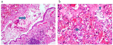
    Figure 7: Foreign body aspiration (arrow), polymorphonuclear leukocytes and erythrocytes are observed in the bronchial lung sections ((H&E X 100).
b: Polymorphonuclear leukocytes, mononuclear inflammatory cells, edema, pneumonia symptoms including erythrocytes (star) and foreign body aspiration (arrow
head) is observed in the alveolar spaces of the lung parenchyma.
As a result, in addition to the damage in the central nervous system and heart, very severe damage also occurs in the lungs and various clinical conditions such as edema, congestion and pneumonia can be seen. All these histopathologic findings may be helpful to consider planning of acute and long term treatment of CO poisoning.
References
- Forbes WH, Sargent F, Roughton FJW. Rate of carbon monoxide uptake by normal men. Am J Physiol. 1945; 143: 594-608.
- Farrow JR, Davis GJ, Roy TM, McCloud LC, Nichols GR. Fetal death due to nonlethal maternal carbon monoxide poisoning. J Forensic Sci. 1990; 35: 1448-1452.
- Hausberg M, Somers VK. Neural circulatory responses to carbon monoxide in healthy humans. Hypertension. 1997; 29: 1114-1118.
- Hee J, Callais F, Momas I, Laurent AM, Min S, Molinier P, et al. Smokers' behaviour and exposure according to cigarette yield and smoking experience. Pharmacol Biochem Behav. 1995; 52: 195-203.
- Yardan T, Cevik Y, Donderici O, Kavalci C, Yilmaz FM, Yilmaz G, et al. Elevated serum S100B protein and neuron-specific enolase levels in carbon monoxide poisoning. Am J Emerg Med. 2009; 27: 838-842.
- Cakir Z, Aslan S, Umudum Z, Acemoglu H, Akoz A, Turkyilmaz S, et al. S-100beta and neuron-specific enolase levels in carbon monoxide-related brain injury. Am J Emerg Med. 2010; 28: 61-67.
- Karapirli M, Kandemir E, Akyol S, Kantarci MN, Kaya M, Akyol O. Forensic and clinical carbon monoxide (CO) poisonings in Turkey: A detailed analysis. J Forensic Leg Med. 2013; 20: 95-101.
- Fedakar R, Turkmen N. Fatal poisonings in the south Marmara region of Turkey, 1996-2003. Eur J Gen Med. 2008; 5: 1-8.
- Karaarslan B, Karapirli M, Kandemir E, Kucuker H, Gurler M, Ince CH, et al. The fatal poisoning pattern of Ankara (Turkey) and nearby cities from 2007 to June 2011: a retrospective study in forensic autopsies. J Forensic Sci. 2013; 58: 1563-1567.
- Uysal C, Celik S, Duzgun Altuntas A, Kandemir E, Kaya M, Karapirli M, et al. Carbon monoxide-related deaths in Ankara between 2001 and 2011. Inhal Toxicol. 2013; 25: 102-106.
- Kati C, Karakus A, Altuntas M, Duran L, lkaya F, Kaya C, et al. Evaluation of acute poisonings in geriatric patients attended to a university emergency clinic. Turkish Journal of Geriatrics. 2013; 16: 286-291.
- Jaffe FA. Pathogenicity of carbon monoxide. Am J Forensic Med Pathol. 1997; 18: 406-410.
- Zhang J, Piantadosi CA. Mitochondrial oxidative stress after carbon monoxide hypoxia in the rat brain. J Clin Invest. 1992; 90: 1193-1199.
- Akyol S, Erdogan S, Idiz N, Celik S, Kaya M, Ucar F, et al. The role of reactive oxygen species and oxidative stress in carbon monoxide toxicity: an in-depth analysis. Redox Rep. 2014; 19: 180-189.
- Chamberland DL, Wilson BD, Weaver LK. Transient cardiac dysfunction in acute carbon monoxide poisoning. Am J Med. 2004; 117: 623-625.
- Brunssen SH, Morgan DL, Parham FM, Harry GJ. Carbon monoxide neurotoxicity: transient inhibition of avoidance response and delayed microglia reaction in the absence of neuronal death. Toxicology. 2003; 194: 51-63.
- Fein A, Grossman RF, Jones JG, Hoeffel J, McKay D. Carbon monoxide effect on alveolar epithelial permeability. Chest. 1980; 78: 726-731.
- Kittredge RD. Pulmonary edema in acute carbon monoxide poisoning. Am J Roentgenol Radium Ther Nucl Med. 1971; 113: 680-681.
- Choi SA, Choi IS. Clinical manifestation and complications in carbon monoxide intoxication. J Korean Neurol Assoc. 1998; 16: 500-505.
- Akdemir HU, Yardan T, Kati C, Duran L, Alacam H, Yavuz Y, et al. The role of S100B protein, neuron-specific enolase, and glial fibrillary acidic protein in the evaluation of hypoxic brain injury in acute carbon monoxide poisoning. Hum Exp Toxicol. 2014; 33: 1113-1120.
- Abelsohn A, Sanborn MD, Jessiman BJ, Weir E. Identifying and managing adverse environmental health effects: 6. Carbon monoxide poisoning. CMAJ. 2002; 166: 1685-1690.
- Bauer I, Pannen BH. Bench-to-bedside review: Carbon monoxide--from mitochondrial poisoning to therapeutic use. Crit Care. 2009; 13: 220.
- McBay AJ. Toxicological findings in fatal poisonings. Clin Chem. 1973; 19: 361-365.
- Thom SR. Dehydrogenase conversion to oxidase and lipid peroxidation in brain after carbon monoxide poisoning. J Appl Physiol (1985). 1992; 73: 1584-1589.
- Raub JA, Mathieu-Nolf M, Hampson NB, Thom SR. Carbon monoxide poisoning--a public health perspective. Toxicology. 2000; 145: 1-14.
- Satran D, Henry CR, Adkinson C, Nicholson CI, Bracha Y, Henry TD. Cardiovascular manifestations of moderate to severe carbon monoxide poisoning. J Am Coll Cardiol. 2005; 45: 1513-1516.
- Cha YS, Cha KC, Kim OH, Lee KH, Hwang SO, Kim H. Features and predictors of myocardial injury in carbon monoxide poisoned patients. Emerg Med J. 2014; 31: 210-215.
- KORB G. [Light and fluorescence microscopic findings on the myocardium in acute poisoning with illumination gas]. Dtsch Z Gesamte Gerichtl Med. 1962; 52: 357-368.
- Fein A, Grossman RF, Jones JG, Hoeffel J, McKay D. Carbon monoxide effect on alveolar epithelial permeability. Chest. 1980; 78: 726-731.
- Sone S, Higashihara T, Kotake T, Morimoto S, Miura T. Pulmonary manifestations in acute carbon monoxide poisoning. Am J Roentgenol Radium Ther Nucl Med. 1974; 120: 865-871.
- Shafer N, Smilay MG, Macmillan FP. Primary myocardial disease in man resulting from acute carbon monoxide poisoning. Am j med. 1965; 38: 316-320.
- Finck PA. Exposure to carbon monoxide: review of the literature and 567 autopsies. Mil Med. 1966; 131: 1513-1539.