
1Department of Ophthalmology of Central Lisbon Hospital Center, Portugal
2Faculty of Medical Sciences, New University of Lisbon, Portugal
*Corresponding author: Dias-Santos A, Departamento de Oftalmologia, Hospital de Santo António dos Capuchos, Alameda de Santo António dos Capuchos, 1169-050 Lisboa, Portugal
Received: October 28, 2014; Accepted: November 22, 2014; Published: November 28, 2014
Citation: Dias-Santos A, Ferreira J, Rosa R, Brito T, Amado D, Cunha JP. The Role of Optical Coherence Tomography in Intracranial Tumor in a Child – Case Report. Austin J Clin Ophthalmol. 2014;1(8): 1037. ISSN : 2381-9162
We report the case of a three-year-old child referred to the Neuro-ophthalmology Department with the diagnosis of benign intracranial tumor in close relation to the left optic nerve and to the optic chiasm. Visual acuity, intrinsic and extrinsic ocular motility and fundoscopy were normal and without asymmetries. Given the age of the child and the impossibility of performing perimetry, we decided to perform visual evoked potentials and Optical Coherence Tomography (OCT) of the optic nerve, with measurement of the peripapillary retinal nerve fiber layer. The result was considered normal and symmetrical. The tomographic study the optic nerve, combined with visual evoked potentials, may be a good co adjuvant in the decision of the appropriate therapeutic approach to benign intracranial masses and in determining the visual prognosis of patients with this type of pathology.
Keywords: Peripapillary retinal nerve fiber layer thickness; Children; Visual evoked potentials; Optical coherence tomography; Intracranial tumors
Arachnoid cysts are duplications or splittings of the arachnoid layer filled with a fluid that is similar to cerebrospinal fluid. They can be either sporadic or secondary to other malformations or diseases and probably represent a congenital defect of the arachnoid layer. Their course is variable as they can develop de novo, grow or decrease in size. Many arachnoid cysts are asymptomatic and can be diagnosed in infancy, childhood or adulthood [1]. The clinical manifestations are dependent on the size and structures compressed by these tumors.
When they are close to the optic pathway, the ophthalmologist is often asked to access its integrity. Clinical evaluation of compressive neuropathies usually involves a functional study and a structural analysis of the Optic Disc (OD). The functional study involves the assessment of visual acuity, color vision, contrast sensitivity, brightness sensitivity, Afferent Pupillary Defect (APD), visual evoked potentials and visual field examination. The structural study involves the analysis of Retinal Nerve Fiber Layer (RNFL) with red-free light, the evaluation of OD pallor and/or excavation and RNFL thickness measurement by Optical Coherence Tomography (OCT).
In non-cooperative patients, the evaluation is limited to the structural analysis of the OD, visual evoked potentials and the test of pupillary reflexes.
A three-year-old Caucasian male presented with a left lateral orbital mass which was painless, non-adherent and had a firm consistency. On examination his visual acuity was 6/6 without correction in both eyes, he had no afferent papillary defect and extrinsic ocular motility was preserved. Biomicroscopy and fundoscopy were also normal as well as the neurological exam (Figure 1). Magnetic Resonance Imaging (MRI) was performed to access the internal extension of the orbital mass. This exam not only confirmed the diagnosis of a left lateral epidermoid cyst but also documented a large left arachnoid cyst occupying the middle cranial fossa. It measured approximately 7.5 x 6.0 x 7.0 centimeters and was slightly pushing the left optic nerve and the optic chiasm (Figure 2).
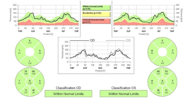
The child was referred to the Neurosurgery Department. They considered that the indication for surgery was based on the effects of the tumor on the optic pathway. Given the impossibility to perform perimetry in this child, we decided to perform visual evoked potentials as well as OCT (Heidelberg Spectralis® Tracking Laser Tomography) of the optic nerve with RNFL thickness measurement (program “RNFL Single Exam Report OU with FoDi™”). Visual evoked potentials proved normal bilaterally. Optic nerve OCT revealed symmetrical optic nerve excavation and peripapillary retinal nerve fiber layer (Figure 3). The Neurosurgery Department thus decided to adopt a conservatory approach based on a regular observation.
Optic nerve excavation and RNFL thickness analysis with OCT is a proven method to access retinal ganglion cell loss as a result of compression of the anterior visual pathway [2-4]. A recent study by Danesh-Meyer et al. showed a strong correlation between standard automated perimetry and RNFL thickness measurement with OCT for compressive optic neuropathies, both topographically and in terms of gravity [5]. This correlation was stronger for the temporal quadrants of the optic disc. They also found a relationship between temporal RNFL thickness loss and visual acuity and color vision. Despite the paucity of normative data for RNFL thickness in children younger than 5 years old [6,7], we believe that symmetry evaluation can be an important diagnostic coadjuvant when combined with visual evoked potentials in the decision of the appropriate therapeutic approach of benign intracranial masses and in determining the visual prognosis of children with this type of pathology.
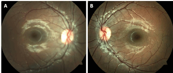
Right eye (A) and left eye (B) retinography was normal.
MRI showing a left lateral epidermoid cyst (wide arrow) and a large left arachnoid cyst (thin arrow).

Optic nerve optical coherence tomography showing symmetrical RNFL thickness in both eyes.
Austin Publishing Group is an emerging open access publisher specialising in Science, Technology and Medicine is dedicated to serve the biomedical community through its initiatives. Austin Publishing Group is an academic publisher with 100+ peer reviewed open access journals in various subjects such as biomedical, Pharma, Life Sciences, Environmental, Engineering and Management. Austin Publishing Group publishes Open Access eBooks providing free access to vast scientific literature.