Case Report
Austin J Clin Pathol. 2014;1(3): 1011.
Extensive Urinary Malakoplakia with Lymph Node Involvement: A Case Report
Yihong Ma1, Jeffrey W Nix2, Ryan C Telford3, Gene Siegal1 and Dejun Shen1*
1Department of Pathology, University of Alabama, USA
2Department of Urology, University of Alabama, USA
3Department of Radiology, University of Alabama, USA
*Corresponding author: Dejun Shen, Department of Pathology, University of Alabama at Birmingham, 619, 19th Street South, NP 3546, Birmingham, AL 35249, USA
Received: May 25, 2014; Accepted: June 24, 2014; Published: June 27, 2014
Abstract
Malakoplakia is an unusual chronic inflammatory disease found mostly in the urinary system, although involvement of other organ systems has been occasionally reported. Masses, ulcers, plaques or papules are formed along the urinary tract and may represent a diagnostic challenge on cystoscopic or imaging studies. Extensive malakoplakia could be systemic and is often associated with immune suppression. We herein report a48-year-old woman with extensive malakoplakia involving the left kidney and renal pelvis, the left ureter, the urinary bladder and the perirenal lymph nodes. The patient was a heavy smoker with a recent history of recurrent urinary tract infections, but had no other significant medical history including immune suppression. She initially presented with abdominal pain, nausea, emesis and unintended weight loss. A CT scan demonstrated significant left hydronephrosis caused by mass lesions in her bladder and left distal ureter. Marked retroperitoneal lymphadenopathy was also noted. Transurethral resection of the bladder tumor and a left laparoscopic ureteronephrectomy revealed extensive malakoplakia upon pathological examination. The patient's retroperitoneal lymphadenopathy was improved as assessed by imaging studies after surgery and antibiotics therapy. Malakoplakia should be considered in the differential diagnosis for patients with a history of urinary tract infections presenting with a mass lesion. Early histological diagnosis and prompt antibiotic treatment may be helpful in avoiding disease progression and potential complications.
Keywords: Malakoplakia; Urinary system; Lymph node involvement; Ureteronephrectomy; Transurethral resection of bladder tumor; Immune suppression
Abbreviations
CT: Computed Tomography; H&E: Hematoxylin and Eosin; TURBT: Transurethral Resection of Bladder Tumor
Introduction
Malakoplakia is an unusual chronic inflammatory disease typically found in the urinary system although involvement of other organ systems has been reported [1]. These lesions usually present as masses, ulcers, plaques or papules along the urinary tract, and are frequently mistaken for a neoplasm on cystoscopic imaging studies [2,3]. Extensive malakoplakia may be associated with a history of immune suppression due to concomitant lymphoma, diabetes mellitus, renal transplantation, or long-term therapy with systemic corticosteroids [4]. Alcoholism has also been reported as a possible risk factor [5]. The pathophysiology of malakoplakiais thought to be associated with insufficient killing of bacteria by macrophages where the partially digested bacteria lead to a deposition of iron and calcium [6]. The deposition forms characteristic intra cytoplasmic Michaelis- Gutmann bodies that are typically 1-10μm in diameter, which can be identified by hematoxylin and eosin (H&E) staining of representative tissue sections. In addition, a calcium stain (von Kossa) is much more sensitive in highlighting their presence [1]. The insulting bacteria are Rhodococcusequior Escherichia coli in most cases [1,6]. Treatment options include antibiotics such as quinolone and trimethoprim-sulfamethoxazole against commonly involved pathogens, ascorbic acid and bethanechol to facilitate intracellular digestion of bacteria, and surgical resection in extreme cases [1]. Discontinuation of immunosuppressive medications and prolonged systemic antibiotics therapy are usually needed to effectively treat malakoplakia. While most of the cases may be controlled by the above therapies, the prognosis may not be ideal in some immune suppressed patients [4].
Case Presentation
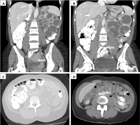
The patient was a 48-year-old African-American woman with a long history of cigarette smoking and a more recent history of recurrent urinary tract infections. No other significant medical history including immune suppression was recorded. She initially presented to an outside hospital with abdominal pain, nausea, emesis, fever and chills. She was diagnosed with and treated for pneumonia at the time. However, she failed to recover from the disease with persistent fever, chills and weight loss. A CT scan showed an enhancing mass along the left bladder base measuring approximately 3.6 cm in greatest dimension (Figure 1A). A synchronous 2.0cm enhancing lesion was also noted within the left distal ureter (Figure 1B) which caused dilatation of the proximal ureter and severe hydro ureteronephrosis (Figure 1A-C), leading to a non-functional left kidney. Marked retroperitoneal lymphadenopathy was also identified, predominantly along the left periaortic space with an index node measuring 2.3cm in greatest diameter (Figure 1C). At the time, a multifocal transitional cell carcinoma with lymph node metastasis was suspected based on the imaging study. A transurethral resection of the bladder tumor (TURBT) was performed and a diagnosis of malakoplakia was established. Meanwhile, a left retrograde pyelogram showed a significant filling defect in the middle to distal ureter. No contrast was seen to advance past this lesion.
Figure 1: Computed tomography scan of the patient with urinary tract malakoplakia. Legend: ACT scan shows left hydronephrosis (A, B and C), a bladder mass lesion (A), a left ureteral mass (B), retroperitoneal lymphadenopathy before (C) and2 months later after clinical treatment (D, 02/23/2014), with the maximal dimension of each being highlighted.
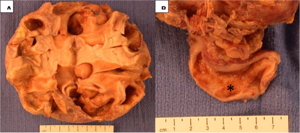
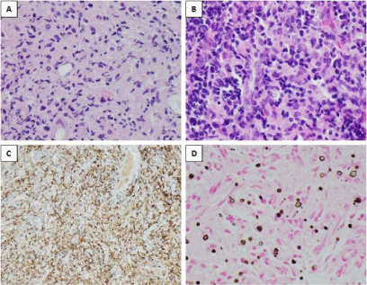
The patient was transferred to our institution for further evaluation and treatment. A repeat TURBT and left ureteral biopsy confirmed the diagnosis of malakoplakia. A left laparoscopic ureteronephrectomy of the non-functioning kidney was performed 3 weeks later. Gross examination showed a massively enlarged kidney with a thinned cortex, dilated calyxes and pelvis and a dilated proximal ureter with a thickened wall (Figure 2). Histological examination revealed extensive malakoplakia involving the entire left kidney including the renal pelvis, the left ureter and multiple grossly evident perirenal lymph nodes on H&E stained sections (Figure 3A and 3B) and by vonkossa stain for calcium (Figure 3D). All the representative histological sections except for the ones from the adrenal gland showed diffuse infiltration with sheets of histiocytes (von hansemann cells), which were highlighted by a CD163 immunostain (Figure 3C). Michaelis-Gutmann bodies were easily identified on the H&E stained sections (Figure 3A and 3B) while the vonkossa stain further highlighted their presence (Figure 3D).
Figure 2: Gross examination of left total ureteronephrectomy specimen. Legend: An enlarged kidney with thinned cortex, dilated calyxes and pelvis (A) and a dilated proximal ureter (*) with a thickened wall (B).
Figure 3: Histopathologic examination of malakoplakia in bladder and lymph nodes. Legend: A. Bladder, H&E, x200; B. Perirenal lymph node, H&E, x200; C. Immunostain for CD163 in the bladder lesion; and D. Von Kosaa stain for calcium insame bladder lesion.
The patient's recovery from her left ureteronephrectomy was complicated by nausea, vomiting, fever, chills and leukocytosis two weeks following surgery, which required another hospitalization. A pelvic abscess was suspected on CT scan although she had no urinary tract symptoms. The patient was treated empirically with antibiotics. She gradually recovered and was doing well at 2 months post nephrectomy follow-up. Her retroperitoneal lymphadenopathy was also improved as assessed by imaging studies (Figure 1C and 1D). She is currently on Levaquin to suppress any residual bacteria.
Discussion
Malakoplakia typically involves the urinary system with symptoms of recurrent infection. Stanton and Maxted based on their literature review found malakoplakia to be located as follows: 58% in the urinary tract (of these 40%in the bladder, 16% in the renal parenchyma, 11% in the ureter and 10% in the renal pelvis); 12% in the gastrointestinal system and 12% in retroperitoneal sites; rare sites made up the remainder which included virtually every organ [1]. Clinical manifestations of malakoplakia remain non-specific. Imaging studies often show plaques and masses, which are frequently misdiagnosed as tumors. Definitive diagnosis is made by histological evaluation. Vonhansemann cells and Michaelis-Gutmann bodies are the two main diagnostic features [1]. Although it is rare, it is known that malakoplakia may be clinically aggressive, which is often associated with underlying immune suppression or some preexisting debilitating conditions [3,5].
In fact, about 50% of reported cases in the literature had associated immune deficiency or autoimmune diseases [4]. Our patient did not demonstrate a significant previous medical history other than recurrent urinary tract infections and heavy smoking. However, a retrospective review of her medical record revealed several abnormal lab values. These included: 1) Three of four "positive" urine cultures for bacteria, even when on antibiotics (bacterial identification was not performed); 2) Anemic with low hemoglobin levels (10-11g/dL), high red blood cell distribution width (20-24) and normal mean cell volume (84-87fl). This type of anemia is typically associated chronic disease; 3) Low serum albumin level (2.9g/dL); and4) Hypomagnesaemia (1.6-1.7mg/dL). Although these abnormal laboratory tests are non-specific, they do suggest that the patient was in a suboptimal health status.
Although the histological diagnosis of malakoplakia was limited to the urinary system and perirenal lymph nodes in this case, the patient may have had more extensive systemic disease or was at least at risk for developing such a condition if left untreated. Our patient's presenting symptoms of nausea; vomiting and abdominal pain raises the possibility of unrecognized gastrointestinal involvement. She also had multiple enlarged retroperitoneal lymph nodes demonstrated by CT scan (Figure 1C). Although these lymph nodes are not biopsied, the index node became smaller after the treatment, suggesting that her retro peritoneal lymphoadenopathy was also caused by malakoplakia.
Conclusion
Malakoplakia should be considered for patients with history of urinary tract infection presenting with a mass. Early histological diagnosis and prompt antibiotic treatment should be helpful in avoiding disease progression and complications.
References
- Stanton MJ, Maxted W. Malacoplakia: a study of the literature and current concepts of pathogenesis, diagnosis and treatment. The Journal of urology. 1981; 125:139-146.
- Ristic-Petrovic A, Stojnev S, Jankovic-Velickovic L, Marjanovic G. Malakoplakia mimics urinary bladder cancer: a case report. Vojnosanitetski pregled Military-medical and pharmaceutical review. 2013; 70: 606-608.
- Abolhasani M, Jafari AM, Asgari M, Salimi H. Renal malakoplakia presenting as a renal mass in a 55-year-old man: a case report. Journal of medical case reports. 2012; 6: 379.
- Leao CA, Duarte MI, Gamba C, Ramos JF, Rossi F, Galvao MM, et al. Malakoplakia after renal transplantation in the current era of immunosuppressive therapy: case report and literature review. Transplant infectious disease : an official journal of the Transplantation Society. 2012; 14: E137-141.
- Ayllon J, Verkarre V, Scotte F, Fournier L, Correas JM, Mejean A, et al. Renal malacoplakia: Case report of a differential diagnosis for renal cell carcinoma. The American journal of case reports. 2012; 13: 38-40.
- Yuoh G, Hove MG, Wen J, Haque AK. Pulmonary malakoplakia in acquired immunodeficiency syndrome: an ultrastructural study of morphogenesis of Michaelis-Gutmann bodies. Modern pathology : an official journal of the United States and Canadian Academy of Pathology, Inc. 1996; 9: 476-483.


