
1Department of Anatomic Pathology, Keelung Chang-Gung Memorial Hospital, Taiwan
2Department of Nephrology, Howard County General Hospital, USA
3Department of Pathology, Johns Hopkins University School of Medicine, US
*Corresponding author: Arend LJ, Department of Pathology, Johns Hopkins University School of Medicine, 720 Rutland Ave., Ross 632E, Baltimore, MD 21205, USA
Received: September 16, 2014; Accepted: October 14, 2014; Published: October 17, 2014
Citation: Chien HP, Liu J and Arend LJ. Acute Tubular Injury in a Woman with Placental Abruption. Austin J Clin Pathol. 2014;1(4): 1019. ISSN : 2381-9170
Here we report the case of a young woman who developed acute renal failure following prolonged cesarean section delivery. The surgical procedure was complicated by placental abruption and uterine leiomyomata. A kidney biopsy for persistent renal failure revealed acute tubular injury from an unexpected source.
Keywords: Placental abruption; Rhabdomyolysis; Myoglobulinuria; Acute tubular injury
A 38-year-old G4P2 woman with no past medical history presented in labor at 38 weeks gestation. She had an uneventful course of pregnancy. There was no history of gestational diabetes, proteinuria, or hypertension. Initial serum creatinine was 1.1mg/dL. Fetal heart rate decelerations were noted and emergency C-section was performed. During the operation, placental abruption was noted. A living and healthy infant was delivered. However, the patient was noted to have several sites of uterine bleeding, which were sutured. Uterine fibroids were also noted. After abdominal closure, massive vaginal bleeding was noted and the patient’s blood pressure dropped. Hysterectomy and left salpingo-oophorectomy was performed. Both ureters were examined prior to closure and found to be intact. Bloody urine was also observed during the operation. After surgery, there was acute renal failure, with creatinine rising from 2.5mg/dL on the first post-op day to 7.2mg/dL over three days. Under the impression of acute tubular necrosis or rapidly progressive glomerulonephritis, a renal biopsy was performed.
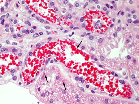
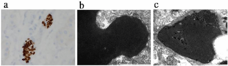
The renal biopsy revealed acute tubular injury with cytoplasmic vacuolization, apical blebbing, focal epithelial flattening, and large intraluminal collections of eosinophilic granular material. Occasional intracytoplasmic eosinophilic globules were present (Figure 1). The intraluminal material was strongly positive for myoglobin (Figure 2a). Ultrastructurally, the renal tubules showed intraluminal electron-dense material with peripheral lucent rims (Figure 2b). Enlarged mitochondria were also noted in the tubular epithelial cells (Figure 2c).
Placental abruption refers to premature placental separation from the uterus prior to the delivery of the fetus. It can cause massive bleeding from the abruption site, disseminated intravascular coagulation, hypertonic uterine contracture, and both maternal and perinatal morbidity. While in most cases the etiology is unclear, placental abruption could be associated with abdominal trauma, uterine anomaly, preeclampsia, and smoking. The patient had multiple uterine leiomyoma with the largest one more than 8cm in diameter. In one retrospective study, multiple and large (>5cm in diameter) uterine leiomyomata are associated with a higher risk of preterm premature rupture of membrane, but no correlation with placental abruption [1].
During the surgery, the patient had massive blood loss (total blood loss approximately four liters) and she developed acute kidney injury after the surgery. Kidney biopsy showed acute tubular injury with cytoplasmic vacuolization and regenerative nuclear changes of tubular cells, and abundant intratubular myoglobin casts. Changes were compatible with myoglobinuric acute tubular injury.
Myoglobinuria, i.e., increased myoglobin in the urine, results from rhabdomyolysis caused by skeletal muscle injury. Causes of rhabdomyolysis include a wide range of mechanical, pharmacologic, metabolic, infectious, and genetic factors [2]. Lampley et al. reported a pregnant woman who used cocaine and 18 hours thereafter presented with placental abruption and rhabdomyolysis, both of which are complications of cocaine use [3]. There was no history of drug abuse in the current patient. The patient received prolonged laparotomy surgery. A prolonged surgery with persistent pressure on skeletal muscle is known to be a cause of postoperative rhabdomyolysis [4]. Other risk factors of postoperative rhabdomyolysis include morbid obesity, diabetes mellitus [5], and certain anesthetic medications, such as propofol [6] and suxamethonium (succinylcholine) [7]. In this patient, the combination of the length of the operation and hemodynamic change might have contributed to the development of myoglobinuric acute tubular injury.
Megamitochondria are a non-specific morphologic change and can be seen in a variety of conditions. The most common causes are ischemia and various types of acute tubular injury [8,9], which are relevant in this case. The megamitochondria observed in acute tubular injury tend to show expansion of mitochondrial matrix with broken cristae [9]. However, in our case, the enlarged mitochondria display normal electron density and well preserved cristae. Therefore, other possible causes of megamitochondria cannot be excluded, including drugs and toxin exposure (eg, antiviral drugs, antiepileptic drugs), diabetes, Wilson’s disease and mitochondriopathies.
After renal biopsy, the patient’s serum creatinine continued to rise, up to 8.9 mg/dL and she was oliguric despite hydration for the suspicion of acute kidney injury from hemodynamic change. After hemodialysis for 2 weeks, the patient was discharged with a stable creatinine at 4.0 mg/dL. One month later, her kidney function improved and serum creatinine was 1.2 mg/dL.
Black arrows indicate intraluminal eosinophilic granules and intracytoplasmic eosinophilic globules.
(a) Myoglobin immunostain: The intratubular granular materials arestrongly positive for myoglobin.
(b) Electron microscopy: Tubular cast showing lamellated inner electronlucentand outer electron-dense rims.
(c) Electron microscopy: Tubular cells contain enlarged and abnormalshaped mitochondria.
Austin Publishing Group is an emerging open access publisher specialising in Science, Technology and Medicine is dedicated to serve the biomedical community through its initiatives. Austin Publishing Group is an academic publisher with 100+ peer reviewed open access journals in various subjects such as biomedical, Pharma, Life Sciences, Environmental, Engineering and Management. Austin Publishing Group publishes Open Access eBooks providing free access to vast scientific literature.