Case Report
Austin J Clin Pathol. 2015;2(1): 1022.
Simultaneous Ipsilateral Presentation of Papillary Renal Cell Carcinoma and Transitional Cell Carcinoma in a Patient With a History of Ductal Adenocarcinoma in Choledoc
Kokenek-Unal TD¹*, Arikok AT², Bozkurt IH³, Karaman H¹ and Alper M²
1Department of Pathology, Kayseri Research and Training Hospital, Turkey
2Department of Pathology, Diskapi Yildirim Beyazit Research and Training Hospital, Turkey
3Department of Urology, Diskapi Yildirim Beyazit Research and Training Hospital, Turkey
*Corresponding author: Kokenek-Unal TD, Department of Pathology, Kayseri Research and Training Hospital, Yakut Mah, Akmescid Cd 3850, Sok.3/30 Kocasinan, Kayseri, Turkey
Received: February 02, 2015; Accepted: February 27, 2015; Published: March 02, 2015
Abstract
Simultaneous occurence of renal cell carcinoma and transitional cell carcinoma of the kidney is a rarely reported case in the literature. But appearance of them subsequent to another primary tumor was not reported until now. Herein, we present a 56 years-old male patient with ipsilateral renal cell carcinoma and low grade noninvasive papillary urothelial carcinoma which were hidden by several calculus in imaging modalities in addition to a history of ductal adenocarcinoma in choledoc.
Keywords: Synchronous multiple primary neoplasms; Renal cell carcinoma; Transitional cell carcinoma; Kidney neoplasms
Abbreviations
RCC: Renal Cell Carcinoma; TCC: Transitional Cell Carcinoma; PCNL: Percutaneous Nephrolithotomy; ESWL: Extracorporeal Shock-Wave Lithotripsy; PRCC: Papillary type Renal Cell Carcinoma
Introduction
Renal Cell Carcinoma (RCC) and Transitional Cell Carcinoma (TCC) are the most common malignant neoplasies in urogenital tract, taken individually. However, the coexistence of them in the same patient, ipsilaterally or contralaterally, is an uncommon case. To our knowledge, approximately 60 cases are reported in literature [1-9]. In addition to that, the presence of multiple primary tumors in the same patient is a situation that is more frequently encountered in recent decades.
Herein, we present a case of synchronous RCC and TCC in a patient with multiple stones in various sizes and a few huge staghorn calculi and multiple simple cysts in both kidneys in addition to the clinical history of choledoc adenocarcinoma.
Case Report
A 56 years-old male with a history of left Percutaneous Nephrolithotomy (PCNL) and left Extracorporeal Shock-Wave Lithotripsy (ESWL) was admitted to urology clinic with complaining of left flank pain. Medical history also revealed that he was diagnosed with ductal adnocarcinoma in choledoc region and had a Whipple operation one year ago in another medical center. According to the pathology report, the adenocarcinoma invaded pancreas and peripancreatic adipose tissue. Lymphovascular invasion was remarkable and perineural invasion was diffuse. Physical, radiological and laboratory examination revealed multiple stones in left ureter and so left ureterorenoscopy and left PCNL were performed. After 6 months, the patient was referred with a new onset of hematuria. In medical examination, stones in various sizes were detected in both kidneys. After complete examination right and left PCNL were performed with monthly interval and stones were removed. During left PCNL, besides multiple stones, papillaroid structures were observed in pelvic region and biopsy was taken intraoperatively. Histological examination revealed that papillary mass was low grade noninvasive papillary urothelial carcinoma. The patient underwent a left radical nephrectomy and ureterectomy.
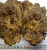
Surgical nephrectomy specimen weighed 335gr and was 16 x 8.5 x 4.5 cm in size with 2.5 cm length urether. On cross section, multiple papillary tumor masses were detected throughout the renal pelvis. Their diameter ranged from 1.5 cm to 4 cm. Besides, in renal cortex a well-defined, cream-yellowish colored, relatively smooth surfaced lesion with diameter of 2.5 cm was observed. Macroscopically, there were also many cysts in cortical region of the kidney, the largest one of 2.5 cm in diameter. Pelvicalyceal system was generally dilated and some calycies were pluged with stones in several sizes (Figure 1).

    Figure 1:  Cross section of resected materyal. A solid lesion in the cortex,
several papillary structures, dilated calyces and stones are easily visible.
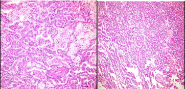
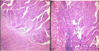
Microscopically, the tumoral mass in renal cortex was diagnosed with papillary renal cell carcinoma (type 1) of Fuhrmann grade 2. It was composed of malignant cells forming papillary structures covered by a single layer of small, hyperchromatic cells with scanty cytoplasm. The tumor papillae contained a delicate fibrovascular core and occasionally foamy macrophages. (Figure 2a & 2b) Renal capsule was not invaded by the tumor. In renal pelvis, another papillary tumor was detected. The tumor was characterized by thin, branching papillary stalks with fibrovascular cores. Variations in nuclear polarity, architecture and size and chromatin content of cells were easily recognized. The nuclei enlarged and showed mild irregular contours. There was no invasion into lamina propria. This tumor was diagnosed as noninvasive papillary urothelial carcinoma. Although it was a low grade in character, it involved almost whole pelvis. (Figure 3a & 3b) Renal vein and resected ureter was not invaded by any of the tumors.

    Figure 2:  Microscopical appearance of solid lesion.
a) Small papillary projections of neoplastic cells.
b) Papillary renal cell carcinoma, papillary structures are filled with
macrophage (arrows). ( H&E, x40, H&E,x 10; a and b respectively)

    Figure 3:  Histological appearance of noninvasive papillary urothelial tumor
of pelvis.
a) Slender papillary stalks with fibrovascular cores. (arrow)
b) Increased stratification and mild pleomorfism of cells are recognized.
(H&E,x10, H&E,x40; a and b respectively)
Discussion
Renal Cell Carcinoma (RCC) is a common neoplasm in general population, but the papillary subtype of RCC (PRCC) is relatively infrequent and constitutes only 10 to 15 % of all RCC cases. Like RCC, PRCC shows slight male predominancy and affects primarily adults [10]. While the most common subtype of renal cell carcinoma was clear cell type in simultaneous RCC and TCC cases [11], our case is the second case in the literature in which the subtype of renal cell carcinoma is papillary type [9].
On the other hand, transitional cell carcinoma is the most common neoplasm in urinary bladder, urether and renal pelvis, even though latter is rarely affected. It occurs mainly in male adults. Although the exact etiology remains unclear, many possible etiologic factors are discussed. Chronic irritation such as inflammation, hydronephrosis, urinary calculi and specific chemicals especially nicotine and analgesics are frequently mentioned [9,11]. Our patient had longstanding many urinary stones in both kidneys. Besides he was a smoker and probably have been using many drugs including powerful analgetics due to the adenocarcinoma of choledoc. In contrast to the most of other reported cases, urothelial carcinoma was a low grade neoplasm and of noninvasive character in our case. In concomitant tumors, as it is pointed out in a study, left kidney is affected 3.2 times more frequently than right kidney [12]. It should be also noted that distinguishing renal tumors of different histology on radiological imaging may be challenging. Even as in our case, radiological and sonographic studies are not able to identify any of the renal tumors due to the existence of multiple and huge calculi and therefore preoperative diagnosis cannot be done.
Furthermore, the prevalence of multiple primary malignancies is between 0.73 % and 11.7 % [13] and most synchronous tumors are seen in genitourinary and gastrointestinal tracts [14,15]. Many studies pointed out that a considerable amount of multiple cancers are related to the kidney tumors [14,15]. Renal cell carcinoma with clear cell histology was encountered the most common type in patients with multiple primary tumors [14]. There are few studies reporting multiple primary tumors in both pancreas and kidney. Some similar genetic mechanisms were identified and coincidence of pancreatic adenocarcinoma and other tumors were expected in especially hereditary cancer syndromes. However, spontaneous occurrence of pancreatic adenocarcinoma and renal cell carcinoma in patients was reported without remarkable history [16]. In addition to that, renal cell carcinoma can metastasize to pancreas and it can cause difficulty in differential diagnosis. Treatment and prognosis of tumors are affected most likely by the more aggressive ones of the multiple tumors.
In conclusion, it should be kept in mind that, kidney is a critical organ which has high coincidence of presence of primary tumors and can be associated with other primary malignancies. Therefore preoperative evaluation must be done carefully.
References
- Fernandez Arjona M, Santos Arrontes D, De Castro Barbosa F, Begara Morillas F, Cortes Aranguez I, Gonzalez L. Synchronous renal clear-cell carcinoma and ipsilateral transitional-cell carcinoma: case report and bibliographic review. Arch Esp Urol. 2005; 58: 460-4653.
- Leveridge M, Isotalo PA, Boang AH, Kawakami J. Synchronous ipsilateral renal cell carcinoma and urothelial carcinoma of the pelvis. CUAJ. 2009; 3: 64-66.
- Chiu CH, Lee CC, Chong PY, Ling CM, Huang HW, Chiang KH, et al. Synchronous ipsilateral renal cell and transitional cell carcinoma. Br J Radiol. 2006; 79: e187-e189.
- Soda T, Nishimura K, Kobayashi Y, Kato T, Tokugawa S, Kishikawa H, et al. A case of synchronous contralateral renal cell carcinoma and urothelial carcinoma. Hinyokika Kiyo. 2009; 5: 491-494.
- Yasuda K, Togo Y, Suzuki T, Yamamoto H, Kokura K. A case of synchronous ipsilateral renal cell carcinoma and renal pelvic urothelial carcinoma (collision tumor): a case report. Hinyokika Kiyo. 2007; 53: 627-630.
- Ulamec M, Stimac G, Kraus O, Kruslin B: Bilateral renal cell carcinoma and concomitant urothelial carcinoma of the renal pelvis and urether: as case report. Lijec Vjesn. 2007; 129: 70-73.
- Hayashi T, Abe T, Nakayama J, Kishikawa H, Sekii K, Yoshioka T, et al. Synchronous ipsilateral renal cell carcinoma and transitional cell carcinoma: report of 2 cases. Hinyokika Kiyo. 2006; 52: 23-26.
- Gomez Garcia I, Rodriguez Patron R, Conde Someso S, Sanz Mayayo E, Garcia Navas R, Palmeiro A. Renal synchronous tumor: association of renal adenocarcinoma and transitional tumor of renal pelvis, in the same kidney, an exceptional discovery. Actas Urol Esp. 2005; 29: 711-714.
- Kirkpantur A, Baydar DE, Altun B, Karcaaltincaba M, Aki T, Yüksel S, et al. Concomitant amyloidosis, renal papillary carcinoma and ipsilateral pelvicalyceal urothelial carcinoma in a patient with familial Mediterranean fever. Amyloid. 2009; 16: 54-59.
- Delahunt B, Eble JN. Papillary renal cell carcinoma. Eble, JN, Sauter G, Epstein, JI., and Sesterhem IA, editors. In: World Health Organisation Classification of Tumours. Pathology and Genetics of Tumours of the Urinary System and Male Genital System. Lyon: IARC Press. 2004; 27-28.
- Holmang S, Johansson SL. Synchronous bilateral ureteral and renal pelvic carcinomas, Incidence, Etiology, Treatment and Outcome. Cancer. 2004; 101: 741-747.
- Hart AP, Brown R, Lechago J, Truong LD. Collision of transitional cell carcinoma and renal cell carcinoma. An immunohistochemical study and review of the literature. Cancer. 1994; 73: 154-159.
- Irimie A, Achimas-Cadariu P, Burz C, Puscas E. Multiple primary malignancies-Epidemiological Analysis at a Single Tertiary Institution. J Gastrointestin Liver Dis. 2010; 19: 69-73.
- Sato S, Shinohara N, Suzuki S, Harabayashi T, Koyanagi T. Multiple primary malignancies in Japanese patients with renal cell carcinoma. Int J of Urol. 2004; 11: 269-275.
- Sato S, Shinohara N, Suzuki S, Harabayashi T, Koyanagi T. Multiple primary malignancies in Japanese patients with renal cell carcinoma. Int J of Urol. 2004; 11: 269-275.
- Beisland C, Talleraas O, Bakke A, Norstein J. Multiple primary malignancies in patients with renal cell carcinoma: a national population-based cohort study. BJU International. 2006; 97: 698-702.
- Müller SA, Pahernik S, Hinz U, Martin DJ, Wente M, Hackert T, et al. Renal tumors and second primary pancreatic tumors: a relationship with clinical impact? Patient Safety in Surgery. 2012; 6: 18.