
Case Report
Austin J Clin Pathol. 2015; 2(2): 1030.
A Non-Healing Ulcer with Underlying Radiologic Findings: A Case of Stewart-Blue Farb Syndrome
Roach NK¹, Chow K² and Lewis M³*
¹Department of Radiology, David Geffen School of Medicine at UCLA, USA
²Department of Radiology, University of Greater Los Angeles Veterans Affairs Healthcare System, USA
³Department of Pathology, University of Greater Los Angeles Veterans Affairs Healthcare System, USA
*Corresponding author: Michael Lewis, Department of Pathology, University of Greater Los Angeles Veterans Affairs Healthcare system, Los Angeles, California, USA
Received: June 23, 2015; Accepted: September 25, 2015; Published: October 09, 2015
Abstract
A 24 year-old man presented with a non-healing, painful ulcer on the dorsum of his left foot. He attributed this to a shrapnel injury to the knee, but further history revealed he had undergone prior intervention for a vascular malformation in that extremity. Radiographs demonstrated soft tissue swelling, vascular calcifications, and osseous remodeling of the second digit. CT demonstrated skin ulceration, soft tissue density at the dorsum of the foot, vascular calcifications, and dilated vasculature as compared to the contralateral side. Acroangiodermatitis describes skin lesions related to underlying vascular malformations, which may be due to a variety of etiologies. Stewart-Bluefarb syndrome denotes acroangiodermatitis related to a congenital arteriovenous malformation. The “pseudo-Kaposi” lesions of Stewart Bluefarb syndrome have similar histopathologic characteristics to Kaposi’s sarcoma, including infiltrating pericytes. Treatment of Stewart-Bluefarb syndrome currently includes embolotherapy and conservative management. As this case demonstrates, prompt diagnosis is crucial for appropriate treatment of this vascular disease.
Keywords: Vascular malformation; Stewart-bluefarb; Amputation
Introduction
Acroangiodermatitis describes a spectrum of skin lesions due to vascular malformations. We report a case of Stewart-Bluefarb syndrome, a rare variant of acroangiodermatitis in which the skin lesions are secondary to underlying congenital arteriovenous malformation [1]. Stewart-Bluefarb syndrome presents in young patients and is an important diagnosis to consider when evaluating a young patient with a lower extremity vascular lesion.
Case Presentation
A 24 year-old male veteran presented with a 5-year history of a non-healing, painful left foot ulcer which he attributed to a training exercise injury. Complications from the ulcer included osteomyelitis requiring antibiotics, multiple debridements’, and skin grafting. His past medical history was significant for a remote history of surgical intervention and endovascular treatment of an Arterial venous Malformation (AVM) of the left lower extremity. Prior pathology specimens from ulcer biopsy revealed non-specific fibro granulation tissue.
Physical examination revealed a 6x2x0.2 cm ulcer on the dorsum of the left foot at the level of the second metatarsal head. The dorsal is pedals and posterior tibia pulses of the left foot were palpable. Arterial duplex ultrasound revealed ankle-brachial indices of 1.2 on the right and 1.0 on the left, with dampened signal in the left political, dorsal is pedals, and posterior tibia arteries. CT angiogram demonstrated occlusion of the left proximal-to-mid political artery with one-vessel posterior tibia artery runoff. Tran cutaneous oxygen measurements across the metatarsal heads and talus ranged from 39-49 mmHg.
Due to his intractable pain and prolonged course of treatment for complications of his ulcer, the patient desired and subsequently underwent trans-metatarsal amputation for definitive treatment.
Pathologic specimen revealed underlying vascular proliferation with cuffs of myopericytes and chronic venous stasis changes of the dermis, suggestive of acroangiodermatitis.
Upon receiving this unexpected diagnosis, further retrospective review of the patient’s history revealed foot radiographs which demonstrated significant calcifications, likely vascular, in the soft tissues of the left foot and lower leg.
Unfortunately, the patient’s course has been complicated by slow wound healing, and he is still undergoing wound care.
Discussion
Acroangiodermatitis describes a spectrum of skin manifestations of underlying vascular malformations. Stewart-Bluefarb syndrome describes skin lesions due to congenital AVM. Other variants of acroangiodermatitis include lesions due to chronic venous stasis (acroangiodermatitis of Mali), due to traumatic or iatrogenic arteriovenous fistula, in the setting of limb paralysis, and at amputation stumps in the setting of suction socket prosthesis use [2-4].
Stewart-Bluefarb syndrome is a rare disorder (fewer than 30 case reports in the literature) first described in 1967 [1]. It is usually diagnosed in patients in their 3rd and 4th decades of life and generally involves a unilateral lower extremity, with preserved palpable distal pulses [5,6]. Hemi-hypertrophy of the affected limb is infrequent, in contradistinction to Klippel-Trenaunay syndrome [4]. The associated skin lesions have been described as “pseudo-Kaposi” in appearance, with reddish-brown or violaceous plaques, papules, and/or nodules [3].
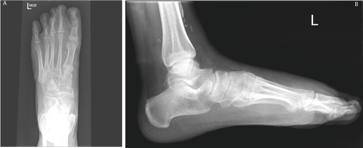
    Figure 1:  Left foot radiographs demonstrate (a) on the PA view, vascular
calcifications between the first and second metatarsals, ankylosis of the
second DIP joint, and bony remodeling of the second middle phalanx,
compatible with a slow, benign adjacent soft tissue process. (b) Lateral
view demonstrates marked soft tissue swelling at the dorsum of the foot and
vascular calcifications of the distal leg.
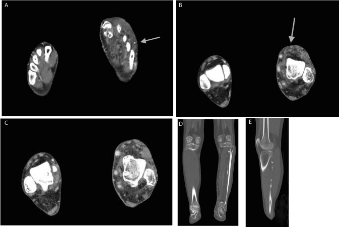
    Figure 2:  CT angiogram (axial) of the bilateral lower extremities demonstrates
soft tissue thickening and stranding of the left foot and ankle. (a) Arrow
indicates region of soft tissue thickening of the dorsum of the left foot and a
vascular calcification at the level of the fourth metatarsal. (b) Arrows indicate
soft tissue thickening and dilatation of vessels at left ankle. (c) Axial images
of the bilateral lower legs demonstrate vascular dilatation and subcutaneous
edema of the left leg as compared to the right. Coronal (d) and sagittal (e)
images of the left leg demonstrate extensive vascular calcifications.
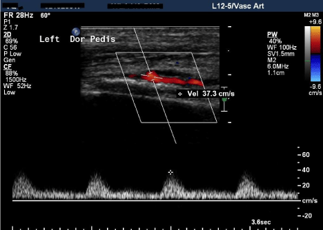
    Figure 3:  Duplex ultrasound of the left foot demonstrates loss of normal
triphasic waveform and diminished amplitude of the dorsal is pedals artery
waveform.
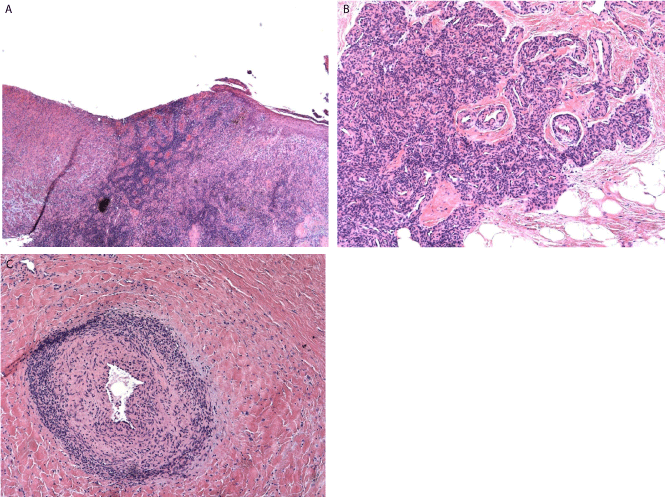
    Figure 4:  Pathological findings from the amputation specimen. (a) This
photomicrograph illustrates skin and subcutaneous tissues near ulcer edge.
Endothelial cell proliferation and angiogenesis are seen in a lobular pattern,
with surrounding pericytes in the dermal layer, more than are normally
present in this region (low magnification; hematoxylin and eosin). (b) Higher
magnification of the region demonstrates pericytes infiltrating capillary walls
and endothelium (hematoxylin and eosin). (c)Near occlusion of this vessel
due to pericyte infiltration of the capillary wall and endothelium causing
luminal narrowing (high magnification; hematoxylin and eosin).
Diagnosis of Stewart-Bluefarb syndrome is based on recognition of dermatologic findings in the setting of congenital AVM. Radiologic findings may be helpful in suggesting the diagnosis.
On plain film radiographs, vascular calcifications may be present. In the case of a large lesion, adjacent bony remodeling, as in this case, may be seen. Focused ultrasound of the vascular malformation may be limited depending size of the lesion, but Doppler interrogation may show high in-flow velocity of the feeding artery and/or arterializations of draining veins [5].
Computed Tomography (CT) angiography demonstrates vascular calcifications and delineates vascular anatomy. Magnetic Resonance Imaging (MRI) assists in characterizing flow pattern and anatomy, including assessing integrity of surrounding fascial planes and involvement of surrounding tissue [3,7]. Digital subtraction angiography is the gold standard for real-time visualization of flow pattern and vessel morphology. Radionuclide quantification of shunting may also be of value [5].
The differential diagnosis for lesions with some or all of these imaging characteristics includes hemangioma, liposarcoma, synovial sarcoma, and malignant familial hyperplasia.
The histopathology features of Stewart-Bluefarb syndrome include angiogenesis with pericytes surrounding and invading vessels. The lesions may be differentiated from Kaposi sarcoma by the absence of “vascular slits” and CD34 expression seen in true Kaposi sarcoma [4,6]. Hemosiderin deposition due to red blood cell extravasation into the dermis may be seen. Prompt diagnosis of acroangiodermatitis is vital for limb preservation, as complications of the syndrome include recurrent infection, hemorrhage, bone demineralization, and poor wound healing [4]. Treatment of small AVMs may include endovascular coiling. AVMs too large or too numerous for successful endovascular therapy may benefit from conservative therapy, including compression stockings and topical steroids [3,6]. Experimental treatments that have been reported include topical glycosaminoglycan and radiation therapy [8,9].
Amputation, as was performed in this case, should be considered a last resort option, as patients with large or multiple AVMs may have reduced wound healing owing to pericyte invasion into vessels and skin. Additionally, AVMs may extend beyond the region of skin findings – as demonstrated in our patient, which likely contributed to his poor wound healing. If amputation is a clinical consideration, detailed attention should be given to vascular imaging findings to ensure that a proposed amputation site is not within a region compromised by vascular malformation. Earlier attention to the possible diagnosis of Stewart-Bluefarb syndrome in this patient may have prompted stronger consideration of more conservative therapy.
References
- Bluefarb, Adams LA. Arteriovenous malformation with angiodermatitis. Arch Derm. 1967; 96: 176.
- Yu GV, Brarebs RM, Vincent AM. Arteriovenous malformation of the foot: a case presentation. J Foot Ankle. 2004; 43: 252.
- Hueso Llombart LB, Alfaro-Rubio A, Serra-Guillen C, Requena C, et al. Stewart-Bluefarb syndrome. 2007; 98: 545.
- Dogan, Boztepe SG, Karaduman A. Pseudo-Kaposi sarcoma: a challenging vascular phenomenon. Dermatol Online J. 2007; 13: 22.
- Rutherford RB. Non invasive evaluation for congenital arteriovenous fistulas and malformations. Semin Vasc Surg. 2012; 25: 49.
- Agrawal S, Rizal A, Agrawal CS, Agrawal A. Pseudo-Kaposi’s sarcoma (Bluefarb-Stewart type). Int J Dermatol. 2005; 44: 136.
- Fayad L, Hazirolan T, Bluemke D, Mitchell S. Vascular malformations in the extremities: emphasis on MR imaging features that guide treatment options. Skeletal Radiol. 2006; 35: 964.
- Hayek S, Atiyeh B, Zgheib E. Stewart-Bluefarb syndrome: review of the literature and case report of chronic ulcer treatment with heparin sulfate. Int Wound J. 2013.
- Goda J, Kapoor R, Yadav BS, Sharma SC. Radiation therapy for intractable bleeding in extremity arteriovenous malformation: considerations on a clinical case. J Med Imaging Radiat Oncol. 2009; 53: 331.