
Case Report
Austin J Clin Pathol. 2015; 2(2): 1031.
A 25-Year-Old Man with Necrotizing Iga Nephropathy and Sub Epithelial Electron Dense Deposits
Fu LY and Moeckel GW*
Department of Pathology, University of Yale, USA
*Corresponding author: Gilbert Moeckel, Department of Pathology, University of Yale, School of Medicine, 310 Cedar Street, LB20, PO: 208023, USA
Received: June 24, 2015; Accepted: September 25, 2015; Published: October 10, 2015
Abstract
IgA Nephropathy (NP) may present with a spectrum of histopathology changes in kidney biopsies, ranging from mild mesangial to diffuse proliferative glomerulonephritis with cellular crescents. IgA deposits are usually present in the mesangium, with additional deposits in the sub endothelial compartment of the capillary loop in some patients with IgA NP. Subepithelial deposits together with fibrinoid necrosis of the glomerulus are uncommon in primary IgA nephropathy. We report the case of a 25-year-old previously healthy man who presented with respiratory symptoms, hematuria and acute kidney injury. A diagnosis of IgA nephropathy was rendered on kidney biopsy based on the presence of mesangial immune complex deposits with dominant IgA immune fluorescence staining. However, instead of the classic glomerular proliferative patterns, this kidney biopsy showed minimal mesangial proliferation with segmental capillary fibrinoid necrosis and sub epithelial electron dense deposits. There was no clinical evidence of Henoch Schoenlein Purpura and the ANCA and Anti-GBM serologies were all negative. In summary we are presenting a kidney biopsy case of unusually active IgA nephropathy, which raises important questions in regard to patient management and clinical outcome.
Keywords: IgA nephropathy; Hematuria; Pathology; Kidney biopsy
Introduction
IgA nephropathy is the most common glomerulonephritis worldwide, and its prevalence is particularly high in South East Asia, where it accounts for 30-50% of renal biopsy diagnosis [1]. Clinical presentations in patients with IgA nephropathy range from asymptomatic microscopic hematuria to rapidly progressive glomerulonephritis to nephrotic syndrome [2-4]. The histopathological spectrum of IgA nephropathy on kidney biopsies is broad, ranging from near normal to severe proliferative glomerulonephritis with cellular crescents, to changes that may resemble primary Focal Segmental Glomerulo Sclerosis (FSGS) [2-5]. Glomerular capillary necrosis is a rare finding in IgA nephropathy and has not been included in the Oxford classification of glomerular lesions, which is used to predict outcome in IgA nephropathy [6-7]. The diagnostic hallmark of IgA nephropathy is the presence of glomerular immune complex deposits with IgA-dominant or co-dominant stain by immunofluorescence. By electron microscopy, electron dense deposits are present particularly in the mesangiumand can extend to the sub endothelial compartment in some cases. Subepithelial electron dense immune complex deposits are a rare finding in IgA nephropathy. We present an unusual case of IgA nephropathy with minimal mesangial proliferation, segmental glomerular capillary necrosis, diffuse interstitial inflammatory infiltrate, tubular necrosis and sub epithelial electron dense deposits.
Case Presentation
Clinical history
A 25 year old man with no significant past medical history presented to emergency department with cough and fever. He reported to have been having a worsening cough for the past 4 days, and the cough was initially dry but became productive of yellow phlegm. He also had fever and chills for the past 2 days, with temperatures measured at home up to 103 F. He had generalized malaise, decreased appetite and poor per oral intake, intermittent nausea and constipation and he complained about voiding dark urine and decreased urine output over the past two days. The remainder physical exam is unremarkable. His primary care physician performed a chest X-ray that showed right lower lobe pneumonia.
In the emergency department, he spiked a fever of 101.2. Repeated chest X-rays reveal elderly pneumonia in the right lower lobe base. Laboratory findings were notable for an elevated keratinize level of 1.6 (previous 1.1 in 2.5 years ago) and normal WBC with left shift (74% neutrophils). Urine analysis showed cloudy urine with 6-10 WBCs, many RBCs, and 2+ proteinuria. He was given a bolus of normal saline, Tylenol and ceftriaxone and admitted for pneumonia and urinary tract infection. A kidney biopsy was performed to evaluate the etiology for acute kidney injury and proteinuria.
Sixteen glomeruli were sample do which three were obsolescent. The open glomeruli appeared enlarged with occasional neutrophils in the glomerular tufts. Six of the glomeruli showed mild increase in mesangial cellularity and matrix deposition. Three out of thirteen glomeruli showed segmental capillary fibrinoid necrosis. The proximal tubules showed acute tubular injury with sloughing of epithelial cells and apical blebbing. Focal tubular necrosis was also noted. The interstitium showed minimal fibrosis in less than 5% of the biopsy tissue. There was diffuse interstitial edema and interstitial infiltrate consisting of lymphocytes, plasma cells and neutrophils with associated tubulitis.
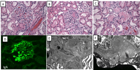
    Figure 1:  Histopathological features of necrotizing IgA nephropathy. (A) Glomerulus shows mild increase in mesanginal cellularity and matrix deposition (400x). (B)
One glomerulus shows segmental capillary necrosis (400x). (C) Focal tubular necrosis in necrotizing IgA nephropathy (400x). (D) Immunofluorescence microscopy
shows 3+ IgA staining in the mesanginal region (400x). (E&F) Ultrastructural examination reveals mesanginal (E) and subepithelial (F) electron dense deposits
(20000x).
By direct immunofluorescence microscopy, there was 3+ IgA in the mesangium and capillary loops. There was 1+ linear accentuation of IgG along the glomerular basement membranes. There was no glomerular, tubular basement membrane, or interstitial staining for IgM, C3, C1q. Fibrinogen showed segmental 2+ positivity in glomerular tufts.
Ultrastructural examination showed segmental corrugation of glomerular basement membranes, segment effacement of foot processes and mild increase in mesangial matrix deposition. Mesangial immune complex deposits and rare sub epithelial immune complex deposits were identified. No sub endothelial immune complex deposits were seen.
Biopsy diagnosis
The biopsy was read as a necrotizing IgA nephropathy.
The necrotizing lesion seen on biopsy raised the question of a possible ANCA-mediated vacuities. Subsequent serological work upturned out negative for ANA, ANCA, dsDNA, and anti-GBM antibodies. Further clinical workup showed no growth in urine and blood cultures, and lower respiratory tract cultures grew normal flora.
The patient was treated for 5 days with antibiotics for possible pneumonia. His temperature returned to normal. He was treated with pulse steroids for 3 days and then transitioned to oral steroids. Serum creatinine levels decreased to 1.2 and proteinuria decreased to 1+ within 1 week post therapy. He started on low dose ACE-I on discharge and was followed up by nephrologist as an outpatient for tapering steroid dose.
Discussion
Biopsy specimens of IgA nephropathy have a range of histological changes [2-5]. The majority show focal or diffuse, mild to severe mesangialhy percellularity often accompanied by an increase in mesangial matrix. Glomeruli might appear near normal early in disease with little proliferative activity. Endocapillary hyper cellularity is observed in around one-third of biopsy samples and is typically focal. Segmental glomerulosclerosis is also a frequent finding in longstanding IgA nephropathies. Extra capillary proliferative lesion, or cellular crescents are present in around one-third of IgA nephropathy biopsies [2]. The crescents are more commonly seen in ANCA–positive IgA nephropathies [8].
Necrotizing lesions are uncommon in primary IgA nephropathy, but are more frequently seen in patients in Henoch-Schonlein Purpura [9]. Glomerular necrosis has not been included in the IgA classifications, which are applied to predict patient outcome [6,7,10].
Patients with necrotic variant of IgA nephropathy exhibit significantly more marked extra capillary proliferation and interstitial accumulation of monocytes and T lymphocytes than seen in our case [11]. Less commonly, capillary necrosis may be the sole feature, presenting either alone or as part of the HSP [12]. These patients did not show significant differences in their clinical presentation compared to those with non-necrotic IgA nephropathy. However, the clinical course in necrotic variant of IgA NP was significant for more frequent flare-ups, and higher rate of progression to end stage renal failure [11].
Another study showed that 5.2%of patients with IgA nephropathy presented with acute tubular necrosis and interstitial nephritis, without specific underlying etiology [13].
IgA deposits are mostly detected in the mesangium but up to one third of patients with IgA NP also show sub endothelial IgA deposits [14,15]. Less commonly, subepithelial deposits are detected [14-16]. It has been suggested that the presence of sub endothelial or sub epithelial deposits is associated with more severe glomerular lesions and higher levels of proteinuria [14,15,17]. Moreover, the presence of IgG positivity has been associated with more aggressive clinical disease and poorer renal outcomes than IgA deposits alone [4].
Our case describes a young patient without the clinical features of HSP, who presented with macroscopic hematuria, sub-nephrotic proteinuria and acute kidney injury. IgA nephropathy was rendered on kidney biopsy due to dominant mesangial IgA staining by direct immune fluorescence and confirmation of mesangial immune complex deposits, as well as subepithelial deposits by EM. Surprisingly, the kidney biopsy showed minimal mesangial proliferation, but segmental capillary necrosis. Moreover, focal tubular necrosis with prominent interstitial inflammatory in filtrates were seen. A possible pauci-immune glomerulonephritis was suspected, but subsequent serologies showed negative ANCA levels.
Post Infectious Glomerulonephritis (PIGN) especially when caused by staphylococcus, may show an IgA dominant immune fluorescence and may mimic IgA nephropathy. In these biopsies also sub epithelial electron dense deposits are identified. However, the PIGN with IgA predominance show usually stronger glomerular endocapillary proliferation and a more prominent neutrophilic glomerular infiltrate. Patients with PIGN also tend to be older and more frequently present with acute renal failure [18].
In conclusion, we present an IgA nephropathy case in a previously healthy young man, whose kidney biopsy showed a few uncommon features including minimal mesangial proliferation with segmental capillary necrosis, focal tubular necrosis, prominent intestinal inflammatory infiltrate, and sub epithelial electron dense deposits. No evidence for ANCA vasculitis, Lupus NP, PIGN or Anti-GBM disease was seen. Our case shows that IgA nephropathy may present as a focally necrotizing GN with minimal proliferation, which may require a more aggressive treatment regimen of the patient.
References
- Schena FP. A retrospective analysis of the natural history of primary IgA nephropathy worldwide. Am J Med. 1990; 89: 209-215.
- Roberts IS. Pathology of IgA nephropathy. Nat Rev Nephrol. 2014; 10: 445-454.
- Wyatt RJ, Julian BA. IgA nephropathy. N Engl J Med. 2013; 368: 2402-2414.
- D'Amico G. Natural history of idiopathicIgA nephropathy: role of clinical and histological prognostic factors. Am J Kidney Dis. 2000; 36: 227-237.
- Ferrario F, Rastaldi MP. Histopathological atlas of renal diseases-IgA nephropathy. J Nephrol. 2004, 17: 351-353.
- Working Group of the International IgA Nephropathy Network and the Renal Pathology Society; Roberts IS, Cook HT, Troyanov S, Alpers CE, Amore A, Barratt J, et al. The Oxford classification of IgA nephropathy: pathology definitions, correlations, and reproducibility. Kidney Int. 2009; 76: 546-556.
- Cattran DC, Coppo R, Cook HT, Feehally J, Roberts IS, Troyanov S, et al. The Oxford classification of IgA nephropathy: rationale, clinicopathological correlations, and classification. Kidney Int. 2009; 76: 534-545.
- Haas M, Jafri J, Bartosh SM, Karp SL, Adler SG, Meehan SM. ANCA-associated crescentic glomerulonephritis with mesangial IgA deposits. Am. J. Kidney Dis. 2000; 36: 709-718.
- Szeto CC, Choi PC, To KF, Li PK, Hui J, Chow KM, et al. Grading of acute and chronic renal lesions in Henoch-Schonleinpurpura. Mod Pathol. 2001; 14: 635-640.
- Jabur WL. Necrotic crescentic glomerulonephritis and IgA nephropathy: Lee-Hass classification revisited. Saudi J Kidney Dis Transpl. 2011; 22: 784-787.
- D'Amico G, Napodano P, Ferrario F, Rastaldi MP, Arrigo G. Idiopathic IgA nephropathy with segmental necrotizing lesions of the capillary wall. Kidney Int. 2001; 59: 682-92.
- Tumlin JA, Madaion MP, Hennigar R. Idiopathic IgA nephropathy: pathogenesis, histopathology, and therapeutic options. Clin J Am SocNephrol. 2007; 2: 1054-1061.
- Mazzarolo-Cruz HM, Penna Dde O, Saldanha LB, Kanashiro EH, Cruz J, Malheiro PS, et al. IgA nephropathy: acute renal failure, acute tubular necrosis, and features of postinfectious acute glomerulonephritis. Ren Fail. 1992; 14: 533-539.
- Lee HS, Choi Y, Lee JS, Yu BH, Koh HI. Ultrastructural changes in IgA nephropathy in relation to histologic and clinical data. Kidney Int. 1989; 35: 880-886.
- Tomino Y, Yagame M, Eguchi K, Miyazaki M, Nomoto Y, Sakai H, et al. Detection of anionic sites and immunoglobulin A deposits in the glomerular capillary walls from patients with IgA nephropathy. J Clin Lab Anal. 1989; 3: 101-107.
- NG WL, Chan KW, Yeung CK, Kwan S. Peripheral glomerular capillary wall lesions in IgA nephropathy and their implications. Pathology. 1984; 16: 324-330.
- Yoshimura M, Kida H, Abe T, Takeda S, Katagiri M, Hattori N. Significance of IgA deposits on the glomerular capillary walls in IgA nephropathy. Am J Kidney Dis. 1987; 9: 404-409.
- Wen YK, Chen ML. Discrimination between post-infectious IgA-dominant glomerulonephritis and idiopathic IgA nephropathy. Renal Failure. 2010; 32: 572-577.