
Case Report
J Dent App. 2014;1(3): 37-39.
Fusion of Mandibular Deciduous Molars: A Case Report
De la Hoz Calvo A1*, Beltri Orta P2 and Chung-Leng Muñoz I3
1Department of Dentistry, European University of Madrid, Spain
2Department of Dentistry, European University of Madrid, Spain
3Department of Dentistry, European University of Madrid, Spain
*Corresponding author: De la Hoz Calvo A, Department of dentistry, European University of Madrid, Calle Tajo, s/n, 28670 Villaviciosa de Odón, Madrid, Spain
Received: June 25, 2014; Accepted: Aug 02, 2014; Published: Aug 04, 2014
Abstract
Tooth fusion is defined as the developmental dental anomaly in which two different tooth buds are fused into one during the development stage. In deciduous dentition, fused teeth occur in 0.1-2.5% of the population, whereas the deciduous molars are the least affected by this anomaly.
This article presents a case report of a 7 year-old male presenting a fusion of the first and second left mandibular deciduous molars. Clinical and radiographic explorations demonstrated no pathology in the deciduous dentition, as well as no involvement of the permanent tooth. As no treatment was necessary for this patient, periodic revisions were scheduled in order to prevent possible root resorption and premature exfoliation of the fused teeth. Additionally, these revisions allowed us to avoid more complex orthodontic problems in the future.
Keywords: Dental fusion; Dental anomaly; Deciduous dentition
Introduction
In the deciduous dentition, as well as in the permanent dentition, anomalies in color, shape, size or number may occur [1].
Fusion is one of the many different types of developmental anomalies. It is defined as the union of two normally separated tooth germs [2]. This anomaly leads to alterations of number, as the union results in one tooth less than normal if the affected tooth is counted as one; size, since the tooth will be much larger as it is the union of two tooth buds, and shape.
Clinically, it may be difficult to differentiate a tooth fusion from another developmental anomaly, such as gemination. Gemination is the attempt of division of a single tooth germ by invagination, resulting in the incomplete formation of two teeth [3]. This particular anomaly does not affect the normal number of teeth, as the affected tooth is counted as one.
Due to this difficulty, the term of "double teeth" is used to designate either the fusion or the gemination [4].
Tooth fusion may occur within the dentin, the enamel, or both. Radiographically, these teeth are observed to have one or more root canals and one or two pulp chambers [5]. On the other hand, geminated teeth present one single root and one root canal [6-8].
This anomaly has a prevalence of 0.1-2.5% in deciduous dentition, and 0.1-0.2% in permanent dentition [5,9-11]. There is an equal distribution between females and males, and there are no significant differences in its location, affecting either maxillary or mandibular arches. Racial differences have been reported to show a higher occurrence in the Japanese population compared to the Caucasian population [9,12,13]. The most affected teeth are the canines and incisors [14,15].
The etiology of tooth fusion is still unknown. Most authors believe that some physical force or pressure provokes the contact between two developing teeth. This contact then produces a necrosis of the epithelial tissue that separates them and leads to their fusion [10,16,17]. Other researchers believe that thalidomide embryopathy, fetal alcohol exposure or hypervitaminosis may be other causes of tooth fusion [10,14,18]. Some also believe there is a genetic foundation for the anomaly, possibly as an autosomal dominant with reduced penetrance [19]. Different studies have reported deciduous double teeth associated with a variety of syndromes, such as Trisomy 21, orodigitofacial syndrome, ectodermal dysplasia or Pierre-Robin syndrome [13].
Since tooth fusion is the union of two tooth buds, there will be anomalies in size and shape, which leads to a number of additional dental problems including aesthetics, spacing, eruption, tooth decay (mainly on the line of fusion), and periodontitis [20-22]. It is also important to emphasize that fused deciduous teeth show a high prevalence (33-70%) of dental agenesis in the permanent dentition [10,18].
No specific treatment is necessary for fused deciduous teeth, although they do require a careful examination. This examination allows for a correct diagnosis and treatment of future problems such as delayed eruption or agenesis of the permanent dentition, crowding or malocclusions [18,19,23].
Case Report
A 7 year-old Caucasian male was examined at the Dental Clinic of the European University of Madrid as part of a general orthodontic evaluation. The patient's medical history was not significant, other than a reported head trauma suffered at age two. The patient's mother stated that she was not aware of similar dental problems within the family. An older brother, who was treated at the same clinic, presented no dental anomalies.
Clinical examination showed that the patient was in early mixed dentition with good oral health and teeth crowding in the right side of the anterior region of the mandible. We observed a fusion of the deciduous left mandibular first and second molars, which resulted in a bigger crown than the contralateral tooth. The number of teeth in the mandible totaled 11, with five deciduous teeth (75-74, 73, 83, 84 and 85) and six permanent teeth (36, 32, 31, 41, 42 and 46) (Figure 1).

Figure 1: Occlusal view of the mandibular arch. Fused lower left primary molars.
The fused tooth presented seven cusps, four buccal and three lingual, separated by occlusal, lingual and buccal grooves. In occlusion, we noted a deviation of the mandibular midline towards the right because of the mesiolingual rotation of the lower right lateral incisor. After measuring the arch length, we found that the left side was shorter than the right side because of the smaller diameter of the fused tooth compared to the sum of the diameter of the two contralateral deciduous molars (Figures 2 and 3).
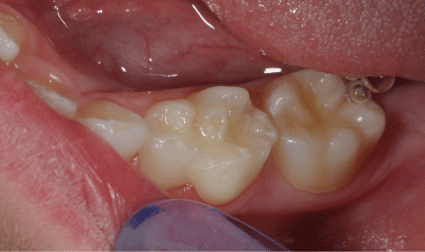
Figure 2: Fused left primary molars in the mandibular arch.
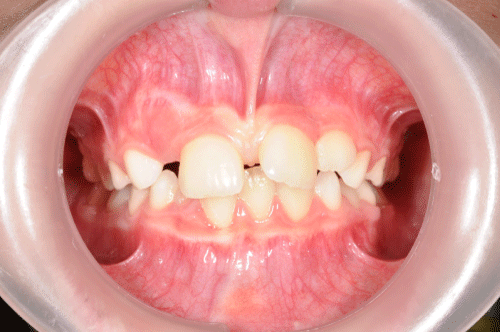
Figure 3: Frontal view in occlusion. Desviation of the midline.
Radiographic examination of the fused teeth showed an enlarged crown with two pulp chambers and three roots. Two permanent bicuspids (34 and 35) could also be observed in the radiographic examination (Figure 4).
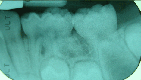
Figure 4: Periapical radiography of fused lower left primary molar. Two permanent bicuspids may be appreciated at the lower part.
Using a panoramic X-ray, it was determined that there was no absence of any permanent teeth.
The child's mother signed an informed consent form prior to photography and radiographic registers.
Discussion
This report demonstrates a rare case of deciduous molars fusion. The clinical examination (crown morphology and number of teeth), as well as the radiographic examination, suggests it is a case of fusion and not germination, as there is one tooth less than normal in the mandible. Morphologically, it is possible to differentiate where the fusion of the two molars occurred.
Dental fusion may be classified as total or partial fusion, depending on the stage in which it happened. A total fusion takes place at the early stages of development before calcification begins, resulting in a normal sized or slightly bigger than normal tooth. If the fusion happens during the calcification stage, the fusion will be partial. In the latter instance, the tooth will have the size of two crowns fused together, a bifid crown, or both [3]. The report presented by Caceda et al. [24] showed a case of fused maxillary molars, in which the fusion was considered to be a complete fusion because the morphology of the crown did not coincide with that of the first and second maxillary deciduous molars. Radiographically, only one pulp chamber could be observed. Conversely, our case could be considered an incomplete fusion, since it is possible to differentiate where the fusion happened between the first and second deciduous molars. Two pulp chambers are clearly perceived in the radiographic examination.
There was no history of previous medical conditions for the patient in this case study, with the exception of a head trauma at age two. However, as the deciduous molar crowns are completely developed and have erupted at this age, we concluded that this trauma did not affect the fusion in any way. Other factors such as hypervitaminosis, fetal alcohol exposure or genetics may have an influence in the appearance of the dental fusion, but there was no clear etiology presents in this case [10,14,18].
Even though dental agenesis of the permanent dentition is common when there is a fusion of the deciduous teeth, all the permanent teeth were present in this case [10,18].
No specific treatment of the fused tooth was necessary at the moment of the patient's examination, as it belonged to the deciduous dentition and had no problems that required dental treatment [18]. Nevertheless, a careful clinical examination and regular dental visits are necessary in order to prevent additional orthodontic problems, particularly considering the fact that the patient already presented space problems in the anterior region of the mandible. Also, the midline was deviated towards the right because of the accentuated rotation of the lower left lateral incisor towards lingual caused by the space problem. Another factor to take into account is the possible eruption problems of the premolars in that region. These eruption problems may come from an incomplete root resorption of the fused molars or the slope towards mesial of the second premolar caused by the lack of space, or both. If this occurs, the extraction of the fused deciduous molars and a complete orthodontic evaluation would be necessary.
Acknowledgment
We would like to thank Dr. Choufani M and Dr. Lopez MC for their help in the elaboration of this case report.
References
- Hamasha AA, Al-Khateeb T. Prevalence of fused and geminated teeth in Jordanian adults. Quintessence Int. 2004; 35: 556-559.
- Chen YH, Cheng NC, Wang YB, Yang CY. Prevalence of congenital dental anomalies in the primary dentition in Taiwan. Pediatr Dent. 2010; 32: 525-529.
- Mohapatra A, Prabhakar AR, Raju OS. An unusual triplication of primary teeth-a rare case report. Quintessence Int. 2010; 41: 815-820.
- Magnússon TE. Hypodontia, hyperdontia, and double formation of primary teeth in Iceland: An epidemiological study. Acta Odontol Scand. 1984; 42: 137-139.
- Demircioglu Guler D, Sen Tunc E, Arici N, Ozkan N. Multidisciplinary management of a fused tooth: a case report. Case Rep Dent. 2013; 2013: 634052.
- Hernandez-Guisado JM, Torres-Lagares D, Infante- Cossio P, Gutierrez-Perez JL. Dental gemination: Report of case. Med Oral. 2002; 7: 231-236.
- Pereira AJA, Fidel RAS, Fidel SR. Maxillary lateral incisor with two root canals: Fusion, germination, or dens invaginatus? Braz Dent J. 2000; 11: 141-146.
- Garattini G, Crozzoli P, Brenna F. Bilateral dental fusion of the upper central incisors: A multidisciplinary approach. J Esthet Dent. 1999; 11: 149-154.
- Aguiló L, Gandia JL, Cibrian R, Catala M. Primary double teeth. A retrospective clinical study of their morphological characteristics and associated anomalies. Int J Paediatr Dent. 1999; 9: 175-183.
- Tuna EB, Yildirim M, Seymen F, Gencay K, Ozgen M. Fused teeth: a review of the treatment options. J Dent Child. 2009; 76: 109-116.
- Ostos Garrido MJ, Peñalva Sanchez MA. Dientes dobles asociados a inclusión dentaria. Posibilidades terapéuticas. Odontología pediátrica. 1996; 5: 91-96.
- Bruce C, Manning-Cox G, Stanback Fryer C, Banks K, Gilliam M. A radiographic survey of dental anomalies in Black pediatric patients. NDA J. 1994; 45: 6-13.
- Boj Quesada JR. Dientes dobles. Arch Odont. 1990; 6: 321-325.
- Modrizuki K, Yoneku T, Yakushiji M, Machida I. The fusion of three primary incisors: report of case. J Dent Child. 1999; 66: 421-425.
- Favalli O, Webb M, Culp J. Bilateral twinning: report of case. ASDC J Dent Child. 1998; 65: 268-271.
- Nunes E, Moraes IG, Novaes PMO, Sousa SMG. Bilateral fusion of mandibular second molars with supernumerary teeth: Case report. Braz Dent J. 2002; 13: 137-141.
- Ballal NV, Kundabala M, Acharya S. Esthetic management of fused carious teeth: A case report. J Esthet Restor Dent. 2006; 18: 13-18.
- Oliván-Rosas G, López -Jiménez J, Giménez-Prats MJ, Piqueras-Hernández M. Considerations and differences in the treatment of a fused tooth. Med Oral. 2004; 9: 224-228.
- Sekerci AE, Sisman Y, Ertas ET, Gümüs H, Ertas H. Clinical and radiographic evaluation and comparison of six cases of fusion involving the primary dentition. J Dent Child. 2012; 79: 34-39.
- Karacay S, Güven G, Koymen R. Management of a fused central incisor in association with a mac- rodont lateral incisor: A case report. Pediatr Dent. 2006; 28: 336-340.
- Malcic A, Prpic-Mehicic G. Conservative treatment of fused teeth in permanent dentition. Acta Stomat Croat. 2005; 39: 327-328.
- Braun A, Appel T, Frentzen M. Endodontic and surgical treatment of a geminated maxillary incisor. Int Endod J. 2003; 36: 380-386.
- Wu CW, Lin YT, Lin YT. Double primary teeth in children under 17 years old and their correlation with permanent successors. Chang Gung Med J. 2010; 33: 188-193.
- Caceda JH, Creath CJ, Thomas JP, Thornton JB. Unilateral fusion of primary molars with the presence of a succedaneous supernumerary tooth: case report. Pediatr Dent. 1994; 16: 53-55.