
Case Report
J Dent App. 2014;1(3): 43-45.
Peripheral Giant Cell Granuloma: a Case Report
Abhishek Kumar1*, Varun Pratap Singh2 and Priyanka Shah3
1Assistant Professor, Department of Pedodontics & Preventive Dentistry, College of Dental Surgery, BP Koirala Institute of Health Sciences, Dharan, Nepal
2Associate Professor & Head, Department of Orthodontics, Nobel Medical College & Teaching Hospital, Biratnagar, Nepal.
3Junior Resident, Department of Pedodontics & Preventive Dentistry, College of Dental Surgery, BP Koirala Institute of Health Sciences, Dharan, Nepal
*Corresponding author: Abhishek Kumar, Assistant Professor, Department of Pedodontics & Preventive Dentistry, College of Dental Surgery, BP Koirala Institute of Health Sciences, Dharan, Nepal
Received: July 12, 2014; Accepted: Aug 16, 2014; Published: Aug 18, 2014
Abstract
Peripheral giant cell granuloma is one of the reactive hyperplastic lesions of the oral cavity, which originates from the periosteum or periodontal membrane following local irritation or chronic trauma. This article reports a case of peripheral giant cell granuloma in an 11 years old female patient. The patient's mother had reported to the Department of Pedodontics and Preventive Dentistry with a chief complaint of swelling in the right back region of upper jaw since four months. Radiographically, (CT scan PNS, maxilla, mandible) revealed ground glass appearance of alveolar process of right maxilla with bony outgrowth along with associated soft tissue component arising from the inner surface of alveolar process of right maxilla.The lesion was excised and sent for histopathologic examination which confirmed the diagnosis of Peripheral Giant Cell Granuloma. The 6-month clinical follow-up revealed uneventful soft issue healing. Early and definite diagnosis correlating clinical, radiologic and histopathologic examination is important for conservative management of such lesion thus eliminating potential risk to adjacent hard tissue structures.
Keywords: Peripheral Giant Cell Granuloma; Giant cells
Introduction
The peripheral giant cell granuloma (PGCG) is a relatively common tumor-like growth of the oral cavity [1] accounting 7% of all benign tumors of the jaw [2]. It is also known as giant cell epulis or peripheral giant cell reparative granuloma [1]. It is not a true neoplasm but rather a benign hyperplastic reactive lesion [3] occurred in response to local irritation such as tooth extraction, inadequate dental restorations, ill-fitting dentures, plaque, calculus, food impaction and chronic trauma [4]. It is more frequent in women than in men, with a slightly higher prevalence in the 30- to 70-year-old-age group, and largely affects the lower jaw (55%) than in the upper jaw [5].
Cases of PGCG have been documented in children, where the lesion appears to be more aggressive, with infiltration of the interproximal crest area, displacement of the adjacent teeth and multiple recurrences [6].
PGCG occurs exclusively on gingiva or edentulous alveolar ridge as variable sized, sessile or pedunculated lesion which is usually deep red to bluish red and bleed easily [7-9]. The final diagnosis however relies on the histological diagnosis [10, 11]. Histologically, fibroblasts are the basic elements. Scattered among the fibroblasts are abundant multinucleated giant cells. Islands of metaplastic bone occasionally may be seen [12]. Other possible sources include osteoblasts, endothelial cells and spindle cells [13].
The purpose of this case report is to illustrate an instance of an aggressive PGCG and to discuss reasonable differential diagnosis, based on the age of the patient history and clinical features.
Case History
An 11-year-old female patient was reported by her mother to the Department of Pedodontics and Preventive Dentistry with a chief complaint of swelling in the right back region of upper jaw since four months. The swelling had reached the present size with no evidence of further growth since past one month. It was associated with pain and difficulty in eating with negative history of trauma or any other associated significant history.
On inspection, the solitary swelling was 2 x1cm in size extending from permanent right maxillary canine to permanent right first molar region. The lesion was sessile, irregular with overlying surface erythematous and ulcerated. On palpation the lesion was soft and tender with tendency to bleed (Figure 1).
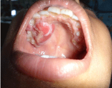
Figure 1: Pre-Operative Intraoral.
Radiologic examination (CT scan, Para Nasal Sinus View (PNS), maxilla, mandible) revealed ground glass appearance of alveolar process of right maxilla with bony outgrowth along with associated soft tissue component arising from the inner surface of alveolar process of right maxilla and also involving interdental gingiva mesial to second premolar in the buccal region (Figure 2). Differential diagnosis of peripheral giant cell granuloma involves giant cell tumour, infantile sarcomas, non-ossifying fibroma which differs from PGCG lesions in consistency and colour; pyogenic granuloma which is difficult to distinguish from PCGC lesions; Cental giant cell granuloma (CGCG) which is an expansive and destructive intraosseous lesion that can perforate the cortex, mimicking PGCG; parulis, which is frequently associated with a necrotic tooth or with periodontal disorder; haemangioma cavernosum, which is distinguished from PGCG lesions by their pulsatile nature; fissured epulis, chondroblastoma which, localized in the gum, may provoke irregular bone destruction below the exophytic lesion; odontogenic cyst.
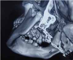
Figure 2: CT Maxilla and Mandible.
Treatment plan included surgical excision of the lesion which was performed under local anaesthesia. There were no complications in the immediate post-operative period (Figure 3). Since PGCGs originate from the periosteum or periodontal membrane following local irritation or chronic trauma, a full mouth scaling was carried out for the patient and oral hygiene instructions were given. No relapse has been observed during the 6 months follow-up (Figure 4).
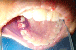
Figure 3: Immediate Post-Operative.
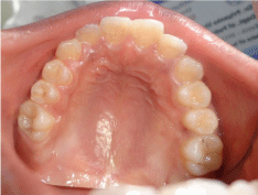
Figure 4: Six months post-operative.
Final diagnosis of Peripheral Giant Cell Granuloma was made based on histopathologic examination which exhibited dense proliferation of osteoclast like giant cells in cellular highly vascularized stroma (Figure 5).
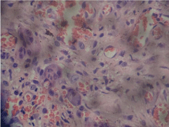
Figure 5: Histopathologic Section.
Discussion
The present report is regarding a case of PGCG successfully treated with excision and curettage. The clinical 6 month follow-up revealed no recurrence and highlighted that the chosen surgical management along with the maintenance of a pertinent oral hygiene are adequate to treat PGCG and prevent its recurrence [1].
In children, reactive oral lesions such as the PGCG can demonstrate a rapid growth and reach significant size within several months of initial diagnosis. These soft tissues nodules may be quite aggressive and resorb bone, interfere with eruption of teeth and produce minor to moderate tooth movement. Radiographs are important for diagnosis to confirm that this giant cell lesion arises within the oral mucosa and does not represent a central bony lesion with cortical perforation and soft tissue extension. Early detection of the PGCG results in more conservative surgery with less risk for tooth and bone loss [14].
Bernier and Cahn proposed the term "Peripheral giant cell reparative granuloma" for the lesion [13]. The latter terminology is currently not being used as the reparative nature of the lesion has not been proved [15]. Today, the term peripheral giant cell granuloma is universally accepted [16].
An origin from periosteum rather than gingiva has been suggested since the lesion can cause superficial erosion of bone and occurs in edentate as well as dentate areas of the jaws [16]. At present, it is generally agreed that PGCG is a reactive, non-neoplastic lesion formed by granuloma-like tissue dominated by multinucleated giant cells [17].
The differential diagnosis of PGCG particularly involves giant cell tumour [3] non-ossifying fibroma which differs from PGCG lesions in consistency and color; pyogenic granuloma which is difficult to distinguish from PCGC lesions; Central Giant Cell Granuloma which is an expansive and destructive intra osseous lesion that can perforate the cortex, mimicking PGCG; chondroblastoma which, localized in the gum, may provoke irregular bone destruction below the exophytic lesion; odontogenic cyst; parulis, which is frequently associated with a necrotic tooth or with periodontal disorder; haemangiomacavernosum, which is distinguished from PGCG lesions by their pulsatile nature; fissured epulis [10]. The peripheral ossifying fibroma is a reactive gingival growth that shares similar clinical features as PGCG. Although this reactive lesion is often ulcerated and inflamed, it lacks the purple or blue discoloration that is commonly associated with PGCG. Identification of small flecks of calcification within the tumescence on a radiograph aids in diagnosing the peripheral ossifying fibroma [17].
The elective treatment is surgical excision with the suppression of the underlying etiologic factors. The periosteum must be included in the excision to prevent recurrences; in fact recurrence is frequent and is observed in 5% and 11% of cases according to Eversole [8] and Mighell [9] respectively. Curettage in addition to the excision to remove the base of the lesion also has been suggested. Recurrence rate of 5.0-70.6% (average 9.9%) has been reported in various epidemiologic studies [8]. This wide variation may be attributed to the surgical technique used in excision [9].
Conclusion
Early and definite diagnosis correlating clinical, radiologic and histopathologic examination is important for conservative management of such lesion thus eliminating potential risk to adjacent hard tissue structures.
References
- Neville BW, Damm DD, Allen CM, Bouquot JE. Soft Tissue Tumors. In Neville BW, Damm DD, Allen CM, Bouquot JE, eds. Oral and Maxillofacial Pathology 3rd ed. St. Louis: Saunders; 2009: 507-563.
- Pour MAH, Rad M, Mojtahedi A. A Survey of Soft tissue Tumor-Like Lesions of Oral Cavity: A Clinicopathological Study. Iran J Pathol. 2008; 3: 81-87.
- Chaparro-Avendano AV, Berini-Aytes L, Gay-Escoda C. Peripheral giant cell granuloma. A report of five cases and reviewof the literature. Med Oral Patol Oral Cir Bucal. 2005; 10: 53-57.
- Motamedi MH, Eshghyar N, Jafari SM, Lassemi E, Navi F, et al. Peripheral and central giant cell granulomas of the jaws: a demographic study. Oral Surg Oral Med Oral Pathol Oral Radiol Endod. 2007; 103: e39-e43.
- Reichart PA, Philipsen H. Atlas de Patología Oral.eds.1 Masson; 2000; 164.
- Wolfson L, Tal H, Covo S. Peripheral giant cell granuloma during orthodontic treatment. Am J Orthod Dentofac Orthop. 96: 519-523.
- Kfir Y, Buchner A, Hansen LS. Reactive lesions of the gingiva. A clinic pathological study of 741 cases. J Periodontol. 1980; 51: 655-661.
- Eversole LR, Rovin S. Reactive lesions of gingiva. J Oral Pathol. 1972; 1: 30-38.
- Reichart PA, Philipsen HP. Gingiva. In Reichart PA, Philipsen HP, eds. Color Atlas of Dental Medicine. Oral Pathology. New York: Thieme; 2000: 148-175.
- Gandara-Rey JM, Pacheo Martina Carneriro JL, Gandara-Vila P, et al. Peripheral giant-cell granuloma: Review of 13 cases. Med Oral. 2002; 7: 254-259.
- Hirshberg A, Kozlovsky A, Schwartz-Arad D, Mardinger O, Kaplan I. Peripheral giant cell granuloma associated with dental implants. J Periodontol. 2003; 74: 1381-1384.
- Regezi JA, Sciubba JJ, Jordan RCK. Red-Blue lesions. In Regezi JA, Sciubba JJ, Jordan RCK, eds. Oral Pathology. Clinical Pathologic Correlations 5th ed. St. Louis: Saunders; 2009: 107-125.
- Rajendran R. Benign and Malignant Tumors of the Oral Cavity. In Rajendran R, Sivapathsundaram B, eds. Shafer's Text book of Oral Pathology 5th ed. New Delhi: Elsevier; 2006: 113-308.
- Flaitz CM. Peripheral giant cell granuloma: a potentially aggressive lesion in children. Pediatr Dent. 2000; 22: 232-233.
- Soames JV, Southam JC. Hyperplastic, neoplastic, and related disorders of oral mucosa. In Soames JV, Southam JC, eds. Oral Pathology 4th ed. New Delhi: Oxford University Press; 2005: 101-115.
- Katsikeris N, Kakarantza-Angelopoulou E, Angelopoulos AP. Peripheral giant cell granuloma. Clinicopathologic study of 224 new cases and review of 956 reported cases. Int J Oral Maxillofac Surg. 1988; 17: 94-99.
- Giansanti JS, Waldron CA. Peripheral giant cell granuloma: review of 720 cases. J Oral Surg 1969; 27: 787-791.