
Case Report
J Dent App. 2014;1(4): 75-77.
Dental Management of Cornelia De Lange Syndrome: Case Report and Literature Review Cornelia De Lange Syndrome
Mariana C Morales-Chávez*
Pediatric Dentist, Magister in Special Care Dentistry. Agreggate Professor and Director of the Dental Research Center, Santa Maria University, Caracas, Venezuela
*Corresponding author: Mariana C Morales-Chávez, Avenida Trinidad con Calle Caracas. Centro Profesional Vizcaya. Piso 3. Oficina 37. Colinas del Tamanaco. Caracas, 1061. Venezuela
Received: June 30, 2014; Accepted: September 16, 2014; Published: September 18, 2014
Abstract
Cornelia de Lange syndrome (CLS) occurs in one out of 10.000 individuals. It is mainly characterized by a delay in growth and development, hirsute, facial dysmorphia, upper-extremity malformation, cardiac defects and dental problems. The aim of this paper is to describe the dental management of a 20 year-old male with CLS, who had multiple dental problems such as caries, periodontal disease, malocclusion and an impacted premolar in the maxilla which produce pain and self-injurious behavior. Physical findings were similar to those previously described by the literature. Dental treatment was carried out under sedation due to the patient's inability to cooperate during dental treatment. After 1 year of follow-up there were no new caries, the periodontal health had improved and the auto aggressive behavior was considerably reduced.
Keywords: Cornelia de Lange syndrome; Brachmann de Lange syndrome; Sedation; Surgical extraction of impact teeth
Introduction
Cornelia de Lange syndrome (CLS) or Brachmann- de Lange syndrome was initially described by Dr. Brachmann in 1916. In 1933 Dr. Cornelia de Lange reported about two cases of this syndrome [1]. CLS is a genetic syndrome which affects between 1/10.000 and 1/60.000 neonates with no racial predilection [2]. It is slightly more common in females (1.3:1).
The genetic and molecular base of this syndrome is not completely clear. However, it is thought to be due to a dominant mutation. A large part of the cases diagnosed as CLS seem to be sporadic and 10% of the cases present chromosomal alterations, translocation of the 3q 26:2-q23 [3-5].
The main clinical characteristics of this syndrome are the delay in growth and development, mental retardation, hirsute, anomalies in the structure of the limbs and distinctive facial characteristics [4].
The facial features are the most clinically consistent and recognizable findings in CLS. Most individuals have microcephaly, short neck, low posterior hairline, hirsute forehead, arched eyebrows and synophrys sometimes with severe ptosis. Thick and long eyelashes, lower ears, flattened midface, short nose and long philtrum are also commonly seen. The oral features include a thin upper lip, a high palate, widely spaced teeth and micrognatia [2,4] Cleft palate is shown by 20% of the patients with this condition [2,6,7].
The extremities are also usually altered by the presence of simian palm groove, micromelia and syndactyly [4]. Ocularly, almost all the patients have problems associated as myopia, ptosis or cronic blefaritis. Medical problems occurred frequently and most often involved the eye and ear, as well as the cardiac and gastrointestinal systems, 25% suffer from congenital heart defects like atrial and ventricular septal defects and hypoplastic left ventricle. These problems and gastrointestinal complications are the main causes of death in patients with CLS [1,2,8].
The syndrome has been associated with certain behavioral and temperamental features such as self-injurious behavior, obsessive-compulsive behaviors, attention deficit disorder with or without hyperactivity, short attention span, depression and autistic features [6,9].
The aim of this study was to identify the principle clinical features of this syndrome in the literature and to describe the dental management of a patient with CLS and how the treatment resulted in the improvement in the quality of life of this patient.
Case Report
The patient was a 20-years-old Venezuelan young male, the first child of a remote consanguineous couple. His younger sister had no signs of dental anomalies. The patient was born at 43 weeks with umbilical cord circumference. During the delivery, the baby inhaled the contaminated amniotic fluid with meconium and experienced respiratory problems. For that reason, the child was hospitalized for 2 weeks. His weight was 2 Kg 400 g and his size 49 cm. The patient was not breastfed. He vomited frequently and at the age of 6 months was diagnosed with severe malnutrition. The cephalic circumference did not grow normally. His genetic evaluation concluded a Cornelia de Lange syndrome. The patient had associated epileptic seizures and underwent treatment with Clonazepam 1.5 mg each day and Phenytoin 8 cc each day.
The patient had short stature. His weight, height and body mass index was very low. He uses a wheelchair because of the psychomotor problems and mental retardation. He also has facial asymmetry, unilateral palpebral ptosis, the eyebrows are very close together (synophrys) and the ears were low implanted (Figure 1).
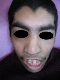
Figure 1: Patient's facial features, which include arched eyebrows and synophrys, severe ptosis, lowest ears, flattened midface and long philtrum.
The oral characteristics were thin upper lip, micrognatia, a high arched and narrow palate and cleft soft palate, a deep overjet, agenesis of superior lateral incisors and a mottled brown pigmentation was present in the enamel and the anterior tooth midline did not coincide (Figure 2). The patient had multiple carious lesions and the marginal gingival was severely swollen inducing a profuse gingival bleeding. The patient's mother informed that he had a severe pain on the right side of the maxilla developing a self-injurious behavior. Clinical and radiographic exams revealed a premolar with horizontal position included in the palatal above (Figure 3).
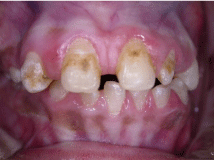
Figure 2: Patient's dental features, which include malocclusion, widely spaced teeth, agenesis of superior lateral incisors and a mottled brown pigmentation in the enamel. The anterior tooth midline does not coincide.
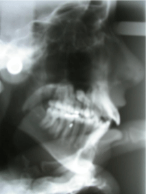
Figure 3: Lateral radiograph of patient showing a premolar with horizontal position included in the palatal above.
Due to difficulties in the patient's behavior and his inability to cooperate during dental treatment because of his mental disability, it was decided that conscious sedation using Midazolam and Propofol would be administered by a pediatric anesthesiologist. All clinical procedures were fully explained to the family, who signed an informed consent form, authorizing treatment.
The included premolar was extracted with a surgical incision and a flat that allowed surgical bucally access. After extraction, the flat was restored to its original position and maintained with suture (Figure 4). All the carious lesions were repaired with composites. Periodontal treatment was carried out with a dental ultrasonic scaler, followed by the application of sealants in the fissures of healthy teeth and fluoride varnish was used on all the teeth. After 2 hours of recovery, detailed oral instructions regarding oral hygiene were given to the patient's family.
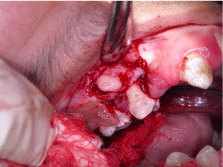
Figure 4: Surgical incision for the extraction of the included premolar.
The patient attended a follow-up session after 1 year. Evidence of improved dental hygiene was observed as no new caries were found. Patient weight increased, self-injurious behavior improved and decrease in his seizures was observed: from a weekly to a monthly episode due to disappearance of pain caused by the included tooth.
Discussion
The Cornelia de Lange syndrome is a rare polimalformation genetic disease. The craniofacial structures are greatly affected. The aforementioned case presents the main characteristics of this syndrome, which have previously been well documented. The patient did not have any cardiac abnormality, which is frequently found in 25% of patients with CLS [6]. The patient experienced seizures and was medicated with Phenytoin and Clonazepam. The neurological alterations appear normally in 23% of patients with CLS [10]. In the anesthetic treatment of a patient with epileptic electroencephalogram waves, even without motor seizure, the problem of the convulsions must receive a very careful attention. Some drugs used in anesthetic procedures like Ketamine were reported to cause convulsions [7]. For such reasons during the treatment of this patient we only used Midazolam and Propofol.
Intraoral findings included a high palate, widely spaced teeth; delayed eruption and cleft soft palate, which often appear in 20% of children with CLS [4].
Oliver et al. [11] reported that self-injurious behavior is frequently identified as part of the behavioral phenotype of Cornelia de Lange syndrome in the 55.6% of the cases. In our patient this was the reason for the consultation and was associated to the included premolar.
The 1-year follow up showed no new caries and an improvement of periodontal health. Also, the patient's mother reported diminished seizure episodes and in the self inflicted behavior. This was probably due to the extraction of the premolar which caused pressure in the right side of the face.
When treating a patient with polimalformative syndrome every precaution must be maximized to ensure that any additional systematic pathology is discarded or detected. It is necessary to request consultations from various specialists in order to evaluate the patient before dental treatment [4].
Conclusion
There are few case histories reported in the literature on the treatment of patients with CLS. We achieved a successful result over a one year period. Treatment of this type of patient should be approached by a team including cardiologist, endocrinologist, pediatric dentist and pediatric anesthesiologist. Dental treatment is restricted by the physical and mental condition of the patient. For this reason a correct evaluation and diagnosis of the real needs of each particular patient is very important.
Why this clinical report is important to pediatric dentists
- This report summarizes key aspects of Cornelia de Lange syndrome and its management.
- The literature review and case report highlight the importance of a complete clinical and radiographic exam to make a correct diagnosis.
- The case report demonstrates that any dental problem can produce behavioral alterations.
Conflict of Interest: There is no conflict of interest.
References
- Jackson L, Kline AD, Barr MA, Koch S. de Lange syndrome: a clinical review of 310 individuals. Am J Med Genet. 1993; 47: 940-946.
- Guadagni MG, Cetrullo N, Piana, G. Cornelia de Lange syndrome: description of the orofacial features and case report. Eur J Paediatr Dent. 2008; 9: 9-13.
- Johns DA, Bhonsale DL, Shivashanker VY, Johns M. Aesthetic and functional management of a patient with Cornelia de Lange syndrome. Contemp Clin Dent.2012; 3: 86-91.
- Grau-Carbó J, López-Jimenez J, Gimenez-Prats MJ, Sánchez-Molins M. Cornelia de Lange syndrome: a case report. Med Oral Patol Oral Cir Bucal. 2007; 12: E445-448.
- Gillis LA, McCallum J, Kaur M, DeScipio C, Yaeger, D, Mariani A, et al. NIPBL mutational analysis in 120 Individuals with Cornelia de Lange syndrome and evaluation of genotipe-phenotype correlations. Am J Hum Genet.2004; 75: 610-623.
- Liu J, Krantz ID. Cornelia de Lange syndrome, cohesin and beyond. Clin Genet. 2009; 76: 303-314.
- Takeshita T, Akita S, Kawahara M. Anesthetic Management of a Patient with Cornelia De Lange syndrome. Anesth Prog.1987; 34: 63-65.
- Noor N, Kazmi Z, Mehnaz A. Cornelia de Lange syndrome. J Coll Physicians Surg Pak. 2012; 22: 412-413.
- Berney TP, Ireland M, Burn J. Behavioural phenotype in Cornelia de Lange syndrome. Arch Dis Child.1999; 81: 333-336.
- Kline AD, Krantz ID, Sommer A, Kliewer M, Jackson LG, FitzPatrick DR, et al. Cornelia de Lange syndrome: clinical review, diagnostic and scoring systems, and anticipatory guidance. Am J Med Genet A. 2007; 143A: 1287-1296.
- Oliver C, Sloneem J, Hall S, Arron K. Self-injurious behaviour in Cornelia de Lange syndrome: 1. Prevalence and phenomenology. J Intellect Disabil Res. 2009; 53: 575-589.