
Case Report
J Dent App. 2014;1(5): 78-80.
Endodontic Management of Fused Teeth: A Case Presentation
Buyukbayram I1, Helvacioglu-Yigit D2 and Ozel E3*
1Acibadem Hospital, Turkey
2Department of Endodontics, University of Kocaeli, Turkey
3Department of Restorative Dentistry, University of Kocaeli, Turkey
*Corresponding author: Ozel E, Department of Restorative Dentistry, University of Kocaeli, Yuvacik, Basiskele, Kocaeli, Turkey
Received: July 26, 2014; Accepted: September 20, 2014; Published: September 22, 2014
Abstract
Fusion is a rare developmental anomaly that is described as a union of two independently developing primary or secondary teeth. It can be either partial or complete, depending on the developmental stage of the teeth when the union occurs, and is infrequently seen in the permanent posterior dentition. Endodontic therapy of these teeth requires special care and careful management because of their abnormal anatomy. This paper reports a case of fusion between a maxillary lateral incisor and a supernumerary tooth and describes the endodontic and restorative management applied.
Keywords: Endodontic therapy; Fusion; Maxillary lateral incisor; Supernumerary tooth
Introduction
Fusion is defined as a union of two separate tooth buds at some stage in their development. The pulp chamber and root canal may be joined or separated, depending on the stage of development at the time of union [1].
Fusion may occur between teeth of the same dentition or between supernumerary teeth. Clinically, a broad crown with a vertical groove extending toward the gingival sulcus is seen. The pulp chamber and the root canals can be joined or separated [2].
The etiology of tooth fusion is still unknown. Genetic predisposition, racial differences, the influence of pressure or physical forces that produce close contact between the developing tooth germs, and trauma have all been attributed as causative factors of tooth fusion [3,4]. Although the etiology is not clear, trauma, disease, or genetics have been suggested as possible causes [5,6].
Most reported cases of fusion are in the incisors and only rarely in the posterior dentition [4,7-10]. Problems with crowding, alignment, and occlusal functions can occur as a result of abnormal morphology and excessive mesiodistal width of a fused molar and a supernumerary tooth [11]. These teeth also tend to be greatly predisposed to caries and periodontal disease [12].
They are joined by the dentine; pulp chambers and root canals may be linked or separated depending on the stage of development at the time of union [13-15].
This paper reports a rare case of a fused maxillary lateral incisor with a supernumerary tooth and treatment with endodontic and restorative treatments.
Case Presentation
A 15-year-old female patient with an esthetic complaint regarding caries was referred to our clinic. Her medical history had no significant data.
The clinical examination revealed that the left maxillary lateral incisor appeared to have an unusual clinical crown and presented as two previously separated crowns joined in the enamel from the incisal of the crown through the cingulum area (Figure 1). A supernumerary tooth was present in the region of the left lateral incisor and seemed to be fused to it. Caries was present along the fissure on the labial surface of the fused teeth. Radiographic examination showed that the lateral incisor and the supernumerary tooth were fused with a separated pulp chamber and two root canals (Figure 2). Mild tenderness on percussion was observed, while there was no tenderness on palpation. The fused teeth were unresponsive to heat, cold, or electrical pulp testing. From the combined clinical and radiographic examinations, a diagnosis of chronic periapical periodontitis was established, and treatment consisted of endodontic therapy and the reconstruction of hard dental tissues with restorative treatment.
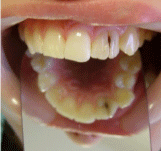
Figure 1: Clinical view of the fused teeth before treatment.
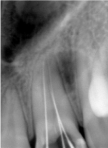
Figure 2: Radiographic view presenting with separated pulp chamber and two root canals.
Two separate anterior access preparations were made in the palatal aspects of each of the clinical crowns. The access preparations became a single cavity after coronal enlargement (Figure 3). Working length was determined by placing files in each canal. The canals were shaped by step-back technique and instrumented through a #35 Headstr�m file (Dentsply Maillefer, Ballaigues, Switzerland), irrigated with 2.5% sodium hypochlorite (Wizard, Rehber Chemistry, Istanbul, Turkey), and dried with sterile paper points. Calcium hydroxide paste (Kalsin, Aktu Tic., Izmir, Turkey) was placed in the canals and temporarily sealed with cotton pellet and zinc phosphate cement. After two weeks the teeth were asymptomatic. The calcium hydroxide paste was removed and obturation was performed using the lateral condensation technique with gutta-percha and AH-Plus sealer (Dentsply, DeTrey, Konstanz, Germany) (Figure 4).
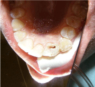
Figure 3: Palatal view of endodontic access cavity.
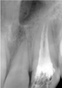
Figure 4: Radiographic view of teeth after treatment.
A two-step self-etch adhesive system (Clearfil SE Bond, Kuraray Medical Inc., Japan) was used in order to perform restorative treatment. The primer was applied to the surface and gently dried with an air syringe for 5 seconds. Adhesive material was then applied to the tooth surface and light-cured for 20 seconds. The teeth were restored with a nanofilled resin composite (CLEARFIL MAJESTY Esthetic, Kuraray Medical Inc., Japan) (Figure 5). All restorative materials were applied according to the manufacturer�s instructions. Finishing and polishing procedures were performed by discs (Sof- Lex, 3M ESPE, St. Paul, MN, USA) and diamond burs (Accurata, Germany), and a second polishing procedure was performed at the one-week recall. One month later, the tooth was asymptomatic and all clinical findings were within normal limits (Figure 6). Selective grinding was performed in order to have a lateral incisor form to the fused teeth. The patient was subsequently lost to follow-up.
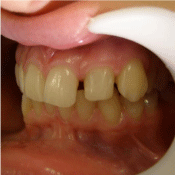
Figure 5: Clinical view of resin composite veneer restoration.
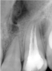
Figure 6: Radiographic view of teeth at the end of one month.
Discussion
Fused teeth can cause various problems, such as caries, periodontal disease, abnormal eruption, impaction, and ectopic eruption of an adjacent tooth [16]. Fused teeth in anterior regions cause severe esthetic problems. Therefore, in the literature, various therapeutic considerations that usually require multidisciplinary treatment steps have been recommended to achieve ideal esthetics and occlusion [17]. In the present case, endodontic and restorative treatment were performed in a multidisciplinary approach.
Supernumerary teeth develop from a third bud arising from the dental lamina near the permanent tooth bud or possibly from splitting of the permanent bud itself [18]. Rare cases of bilateral fusion of maxillary anterior teeth with supernumerary teeth have been reported in which they were treated with nonsurgical endodontic therapy [19].
Fusion is the union of two separate tooth buds in the developmental stage. Gemination and fusion have some similar clinical characteristics. Differential diagnoses can be made according to the number of teeth on the dental arch. In gemination, the arch will contain the normal complement of teeth, whereas in fusion there will be one less than the normal complement. However, it is difficult to differentiate fusion from gemination, especially when these anomalies take place together with hypodontia or a supernumerary tooth [20].
Many different multidisciplinary approaches for the treatment of fused teeth have been suggested because of the considerations of the endodontic, esthetic, orthodontic, periodontal, and functional problems [21]. The endodontic therapy approach depends on whether there are independent pulp chambers and canals or one pulp chamber and two canals. The esthetic criterion is the determining factor when choosing restorative treatment. In the case where the pulp chambers and canals are separated, separation and extraction of the anomalous tooth with orthodontic closing of the space and reshaping of the teeth is one of the treatment options [22]. Other treatments may involve surgical separation with restoration of both teeth [23]. The third possibility stated in the literature is selective grinding of the fused teeth so that the width of the crown is reduced [17]. In this case report, fused teeth were restored together because the abnormal space between the teeth would be difficult to restore esthetically in the case of extraction of the anomalous tooth. Following a resin composite veneer restoration, selective grinding was performed in order to have a lateral incisor form to the fused teeth.
In the current case, the endodontic and restorative treatment of two separated root canals of fused teeth is presented. The root canal treatment and direct resin composite restoration (as a composite veneer) were successfully performed, respectively. As a result of these combined treatments, the patient was pleased with the overall esthetic and functional results.
References
- Duncan WK, Helpin ML. Bilateral fusion and gemination: a literature analysis and case report. Oral Surg, Oral Med, Oral Pathol. 1987; 64: 82-87.
- Tsesis I, Steinbock N, Rosenberg E, Kaufman AY. Endodontic treatment of developmental anomalies in posterior teeth: treatment of geminated/fused teeth - report of two cases. Int Endod J. 2003; 36: 372-379.
- Zeylabi A, Shirani F, Heidari F, Farhad AR. Endodontic management of a fused mandibular third molar and distomolar: a case report. Aust Endod J. 2010; 36: 29-31.
- Kaffe I, Littner MM, Begleiter A, Buchner A. Fusion of permanent molars. Quintessence Int. 1982; 13: 1237-1239.
- Ozalp SO, Tuncer BB, Tulunoglu O, Akkaya S. Endodontic and orthodontic treatment of fused maxillary central incisors: a case report. Dent Traumatol. 2008; 24: e34-37.
- Mancuso A. The treatment of fusion and supernumerary maxillary central incisors: a case report. Gen Dent. 2003; 51: 343-345.
- Peyrano A, Zmener O. Endodontic management of mandibular lateral incisor fused with supernumerary tooth. Endod Dent Traumatol. 1995; 11: 196-198.
- Friedman S, Mor H, Stabholz A. Endodontic therapy of a fused permanent maxillary lateral incisor. J Endod. 1984; 10: 449-451.
- Mader CL. Fusion of teeth. J Am Dent Assoc. 1979; 98: 62-64.
- Goldberg JM, Gross M, Rankow H. Endodontic therapy involving fused mandibular second and third molars. J Endod. 1985; 11: 346-347.
- Ghoddusi J, Zarei M, Jafarzadeh H. Endodontic treatment of a supernumerary tooth fused to a mandibular second molar: a case report. J Oral Sci. 2006; 48: 39-41.
- Ballal S, Sachdeva GS, Kandaswamy D. Endodontic management of a fused mandibular second molar and paramolar with the aid of spiral computed tomography: a case report. J Endod. 2007; 33: 1247-1251.
- Turell IL, Zmener O. Endodontic Therapy in a fused mandibular molar. J Endod. 1999; 25: 208-209.
- Caceda JH, Creath CJ, Thomas JP, Thornton JB. Unilateral fusion of primary molars with the presence of a succedaneous supernumerary tooth: case report. Pediatr Dent. 1994; 16: 53-55.
- Pereira AJA, Fidel RAS, Fidel SR. Maxillary lateral incisor with two root canal: fusion, gemination or dens invaginatus? Brazil Dent J. 2000; 11: 141-146.
- de Siqueira VC, Braga TL, Martins MA, Raitz R, Martins MD. Dental fusion and dens evaginatus in the permanent dentition: literature review and clinical case report with conservative treatment. J Dent Child. 2004; 71: 69-72.
- Karacay S, Gurton U, Olmez H, Koymen G. Multidisciplinary treatment of "twinned" permanent teeth: two case reports. J Dent Child. 2004; 71: 80-86.
- Indra R, Srinivasan MR, Farzana H, Karthikeyan K. Endodontic management of a fused maxillary lateral incisor with a supernumerary tooth: a case report. J Endod. 2006; 32: 1217-1219.
- Nunes E, de Moraes IG, de Novaes PM, de Sousa SM. Bilateral fusion of mandibular second molars with supernumerary teeth: case report. Brazil Dent J. 2002; 13: 137-141.
- Malcic A, Prpic-Mehicic G. Conservative treatment of fused teeth in permanent dentition. Acta Stomatologica Croatia. 2005; 39: 327-328.
- Kim SY, Choi SC, Chung YJ. Management of the fused permanent upper lateral incisor: a case report. Oral Surg Oral Med Oral Pathol Oral Radiol Endod. 2011; 111: 649-652.
- Velasco LF, De Aranjo FB, Ferreira ES, Velasco LE. Esthetic and functional treatment of a fused permanent tooth: a case report. Quintessence Int. 1997; 28: 677-680.
- Hulsmann M, Bahr R, Grohmann U. Hemisection and vital treatment of a fused tooth - literature review and case report. Endod Dent Traumatol. 1997; 13: 253-258.