
Research Article
J Dent App. 2014;1(5): 81-87.
Evaluation of the Correlation between Bacterial Leakage and the Quality of the Filling in Root Canals Obturated by using different Sealer-core Material Combinations
Akman M1*, Eldeniz AU2 and Belli S2
1Department of Endodontics, Necmettin Erbakan University, Konya, Turkey
2Department of Endodontics, Selcuk University, Konya, Turkey
*Corresponding author: Melek Akman, Department of Endodontics, Necmettin Erbakan University, Faculty of Dentistry, Konya, Turkey
Received: August 11, 2014; Accepted: September 20, 2014; Published: September 22, 2014
Abstract
Objective: To evaluate the obturation quality of roots filled with different resin based sealer-core material combinations and to investigate the correlation between quality of the filling and bacterial leakage.
Materials and Methods: Single-rooted human teeth were divided into seven test (n=15) and two control groups (n=12) as follows: HybridRootSEAL/ K3 gutta-percha, HybridRootSEAL/Resilon, RealSeal/Resilon, RealSeal/K3 gutta-percha, AH Plus Jet/K3 gutta-percha, Acroseal/K3 gutta-percha and Diaket/K3 gutta-percha. Leakage was assessed for turbidity of the broth in the lower chamber every day during the test-period of 60 days. Survival analysis was performed (Kaplan-Meier Test). Six roots were then randomly selected from each group and were serially sectioned to obtain fifteen sections from each. Digital images were taken under microscope (50X; LeicaMZ16A) using IC3D camera (Leica). The mean area of the gaps/voids between the sealer-root dentin, sealer-core materials and inside the sealer mass was measured and recorded as μm2. The data was statistically analyzed.
Results: No significant difference was found among the test groups according to their resistance to bacterial leakage (p=0.76). The percentage of non-leaked specimens was highest for the RealSeal/Resilon group (%40). Groups, sections and locations have an effect on the quantity of the gaps/voids (p<0.001). Hybrid Root SEAL/Resilon group showed more gaps/voids when compared with the other groups (p<0.05). The gaps/voids occurred mostly between the core material and sealer (p<0.05).
Conclusions: No correlation was found between the quality of the root filling and bacterial leakage values. Adhesion between the core materials and resin-based root canal sealers still needs to be improved.
Keywords: Bacterial leakage; Gap; Resin based sealer; Single cone technique; Root filling quality
Introduction
One of the requirements for a successful root canal filling is the achievement and maintenance of a tight seal [1] that entombs the remaining microorganism or prevents the ingress of new bacteria and their by-products to the periradicular tissues [2]. Therefore, complete obturation of the pulp space with a core material, which usually is gutta-percha in combination with a root canal sealer, is a standard method of care [3]. In 2003, Resilon (Pentron Clinical Technologies, Wallingford, CT), a thermoplastic filled polymer, has been introduced to the dental market as an alternative to gutta-percha. Resilon contains methacryloxy groups and thus can be used in conjunction with all multi-methacrylate based sealers such as; Epiphany (Pentron Clinical Technologies, Wallingford, CT), RealSeal and RealSeal SE (Sybron Endo, Orange, CA).
A self-adhesive, methacrylate resin-based sealer (Hybrid Root SEAL; Parkell Inc, Farmington, NY) has been introduced commercially [4]. The sealer is self-etching and hydrophilic due to the inclusion of an acidic resin monomer 4-methacryloyloxyethyl trimellitate anhydride and is recommended for use exclusively with either cold-compaction or single-cone techniques [5].
The quality of a root canal filling is highly dependent upon the distribution of the sealer and its adherence to the dentinal walls and gutta-percha [6]. Endodontic sealers are capable of filling imperfections and increasing the adaptation of gutta-percha [7]. On the other hand, root canal sealers are not dimensionally stable and may dissolve partially over time [8,9]. Therefore, to achieve optimal results, it is important to maximize the amount of solid core material and minimize the amount of sealer [10]. Non compaction, the single-cone filling of root canals, has been reviewed [11,12] with the introduction of greater taper master cones that closely match the geometry of nickel-titanium instrumentation systems [13]. Manufacturers claim that the matched taper points can effectively fill tapered canals because they correspond to canal shapes created by similarly tapered instruments [13].
Many studies have been performed to evaluate coronal leakage with various methods until now; dyes, radioisotopes, fluid filtration, electrochemical circuits, endotoxin or bacteria [14]. Using bacteria as a leakage tracer is believed to provide more biologically significant and clinically relevant information [15]. Enteroccocci, especially Enterococcus strains are frequently found in filled root canal systems [16]. The relatively low isolation rate of Enterecocci in primary root canal infections [17] proves that they might colonize the root canal system through coronal leakage after the root filling.
In recent years, the quality of filling techniques and related areas in the gutta-percha region has received increasing attention in studies. This approach was first described by Eguchi et al. [18]. Gaps in gutta-percha sealer-filled areas were measured at the horizontal sections in other similar studies, and the percentages of the filled areas were calculated [10,19]. Previous studies in this field were limited because they only measured and calculated the percentages of the surface area of filling materials and voids by analyzing the sectioned roots and using digital imaging software [11,20,21]. Three-dimensional (3-D) measurements can give more information about the dimensions and the locations of the gaps or voids.
In a previous study, De Deus et al. [22] have evaluated the correlation between canal filled area and bacterial leakage in oval shaped canals and as a result they have found no correlation between the apical seal and the quality of root filling.
The aim of this study was to evaluate the correlation between the bacterial leakage and the quality of the root fillings using a 3-D Topographical Measurement System. Thus the hypothesis tested was that a correlation exists among the bacterial leakage values and the quality of the root fillings.
Materials and Methods
One hundred twenty nine teeth extracted for orthodontic reasons or periodontal problems with similar size and single canals were used after obtaining IRB approval. The crowns were sectioned at the cemento-enamel junction with diamond disc under water cooling. The calculus and soft tissues on the root surfaces were removed with scalpel blades. Working length (WL) was determined by subtracting 1-mm from #10 K-file that was visible at the foramen. All roots were adjusted to 15 ± 0.5-mm in length. The apical part of the root canals was prepared with K3 Ni-Ti rotary instruments (SybronEndo, Orange, CA) to a size of .06/#45. The prepared root samples were randomly divided into 7 experimental groups (15 roots each) and another 2 groups as the positive and negative controls (12 roots each).
The smear layer were removed in an ultrasonic bath (Bandelin Sonorex, Berlin, Germany) using 5.25% NaOCl followed by 17% EDTA and the specimens were rinsed with distilled water for 3 min to remove remnants of these solutions and autoclaved in vials containing distilled water for 20 min at 121°C. Canals dried with sterile paper points (Dentsply-Maillefer, Ballaigues, Switzerland), and filled with five different sealers to create seven test-groups and two control-groups as follows:
Group 1: Hybrid Root SEAL/K3 gutta-percha,
Group 2: Hybrid Root SEAL/Resilon,
Group 3: RealSeal/K3 gutta-percha,
Group 4: RealSeal/Resilon,
Group 5: AH Plus Jet/K3 gutta-percha,
Group 6: Acroseal/K3 gutta-percha,
Group 7: Diaket/K3 gutta-percha.
The sealers were mixed according to the manufacturer's instructions, and applied to the canals with a lentulo spiral (Dentsply, Maileffer, Ballaigues, Switzerland). The root canals were filled with either .06/#45 gutta-percha (K3, SybronEndo, CA, USA) or Resilon (Resilon Research LLC, Madison, CT) cones under aseptic conditions. In RealSeal/Resilon and RealSeal/K3 gutta-percha groups, self-etching primer of the RealSeal system (RealSeal; SybronEndo, Glendora, CA, USA) was applied to the root canal walls with a micro-brush and excess primer was blotted dry with paper points. The teeth in the positive control group were filled with gutta-percha but without sealer. The teeth in the negative control group were also filled with gutta-percha without sealer and completely covered with sticky wax. All experimental, negative and positive control groups were stored under same conditions for 14 days at 37°C and 100% relative humidity to allow enough time to the sealers for complete setting before the evaluation.
Bacterial leakage test
Bacterial leakage was tested using the same model and materials explained by Eldeniz and Ørstavik [23] with using streptomycin resistant Enterococcus faecalis. The mounts were kept at 37°C throughout the experiment for 60 days. The bacteria in the upper chamber were replaced with fresh broth every second day. Bacterial penetration along the root fillings was detected by turbidity observed in the lower chamber. The Kaplan-Meier method was used to construct survival curves for each group and alpha type error set at 0.05. Specimens showing no leakage over the 60 day observation were computed with an event time of 60 days as censored variables.
Scanning electron microscope (SEM) preparation
One specimen with microbial leakage was randomly selected from each group after the completion of the leakage test. The specimen was sectioned horizontally to see dentin-sealer-core interfaces. After fixation with 3% glutaraldehyde (Sigma-Aldrich, St. Louis, MO), they were mounted onto a SEM specimen stub, gold-sputtered (Polaron Sc7620, VG Microtech Inc., Japan) and SEM photomicrographs (Leo 440, Electron Microscopy Ltd.Cambridge, UK) were taken at x500 to x5000 original magnification.
Measurement of the root filling quality
Six roots were randomly selected from each group. Fifteen 0.3 ± 0.02 mm thick sections were obtained under water cooling using Isomet Saw (Isomet, Buehler, Ltd., Lake Bluff, IL, USA) from each root beginning from 1 ± 0.3 mm apically. Stereo digital images were taken from the sections at 50X magnification under a microscope (Leica MZ16A) (Figure 1). A 3-D Topographical Measurement System was used for the quantitative measurement of the voids or gaps in the root fillings. This system consisted of an integrated stereo camera, software and a display system. In order to digitally capture, display, and measure a 3-D object in the accurate fashion, a pair of photos, each with a slightly different perspective of the specimen, needed to be attained. The Leica IC 3D is a digital camera with two independent RGB sensors and captures pairs of stereo images (stereo-pairs) for 3-D analysis. The 3-D reconstruction was based on two images of the specimen taken from slightly different angles. Leica Stereo Explorer software automatically determined which pixels in the two images of the stereo-pair belong together and calculated the topography of the specimen (taking into consideration the parameters of angle and magnification) as a Digital Surface Model (DSM) (Figure 2). This complete 3D data record served as the basis for a variety of different surface analyses.
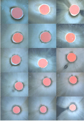
Figure 1: Stereo digital images taken from the sections at 50X magnification under a microscope in RealSeal / K3 gutta- percha group.
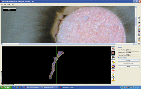
Figure 2: DSM model used for calculation of the mean area of a gap.
Evaluations for the gaps or voids at the sections were done according to the following classifications:
- Voids occurred between the root dentin and sealer,
- Voids occurred between the core and sealer,
- Voids occurred within the sealer mass.
The means of the gap or void areas were measured using 3-D Topographical Measurement System and recorded as μm2. The statistical analyses were performed using SPSS Version 16.0 (α=0.05). Multivariate Analysis of Variance and Tukey HSD tests were performed to examine the effects of groups, sections and regions on the amount of gaps or voids. To evaluate the distribution of the gaps or voids according to the regions or sections, the data were evaluated using One Way ANOVA and Tukey HSD tests.
Results
Bacterial leakage
The survival curves are given in Figure 3. While all positive controls showed turbidity in the lower chambers within 24 to 48 hours, none of the negative controls leaked throughout the entire test period. The resistance of all the tested sealers to the penetration of the bacteria was better when compared with positive control in which no sealer was used (p<0.05). Kaplan-Meier test showed no statistical significant differences between the experimental groups with respect to leakage over time (p = 0.76). All samples which were taken from the bottom chambers after the occurred turbidity demonstrated only the presence of E. faecalis. The percentage of leak-proof specimens was highest for the RealSeal/Resilon group (% 40) and lowest for the AH Plus Jet/K3 gutta-percha group (% 6.7) and were shown in Table 1. Figure 4 shows scanning electron micrographs of horizontally sectioned leaked samples.
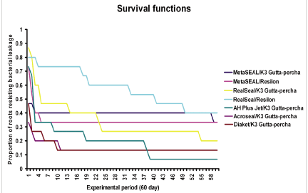
Figure 3: Kaplan Meier survival curve showing the proportion of samples resisting leakage in each experimental group.
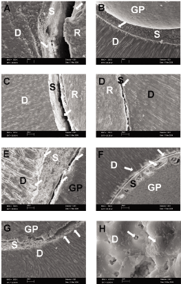
Figure 4: Scanning electron micrographs of horizontally sectioned samples (A) Gaps between Hybrid Root SEAL/Resilon (B) Resin devoid areas between Hybrid Root SEAL/dentin (C) Voids between RealSeal sealer and Resilon (D) Gap formation between RealSeal and dentin (E) Impaired adhesion of AH Plus sealer to dentin and gutta-percha (F) Voids within Acroseal sealer and its debonding from root canal walls forming gaps in some areas (G) Gap formation and debonding of Diaket sealer from dentin and gutta-percha due to the shrinkage (H) E. feacalis penetration and tubule invasion in smear depleted dentin in this bacterial leakage set-up.
![]()
    Group
    Material
    n
    P
    m
    p (%)


    1
    Hybrid Root SEAL/K3    Gutta-percha
    15
    10/15
    1
    33.3%


    2
    Hybrid Root SEAL/Resilon
    15
    10/15
    3
    33.3%


    3
    RealSeal/K3    Gutta-percha
    15
    12/15
    5
    20.0%


    4
    RealSeal/Resilon
    15
    9/15
    41
    40.0%


    5
    AH Plus Jet/K3    Gutta-percha
    15
    14/15
    3
    6.7%


    6
    Acroseal/K3    Gutta-percha
    15
    13/15
    1
    13.3%


    7
    Diaket/K3 Gutta-percha
    15
    13/15
    1
    13.3%


    8
    Negative Control
    12
    0/12
    60
    100%


    9
    Positive Control
    12
    12/12
    0
    0%
Table 1: Bacterial leakage results for the experimental and control groups.
Root filling quality
The distribution of the gaps or voids among the test groups are shown in Table 2. Groups, regions, sections and group-area interactions all have an effect on the amount of gaps or voids (p<0.05). Group-section, region-section, group-section-region interactions did not affect the amount of gaps or voids (p>0.05). Group 2 showed more gaps or voids when compared to the other groups (p<0.05). In all the tested groups, gaps or voids were mostly found between the core material and the sealer (p<0.05). The average amount of gaps or voids was lower at the apical sections when compared to the coronal sections (p<0.05). Gaps/voids occurred mostly between the tested resin based sealers and the core material (p<0.05) except the groups AH Plus Jet/K3 gutta-percha (Figure 5) and Acroseal group (Figure 6). The location of the gaps/voids was mostly within the sealer mass in Acroseal/K3 gutta-percha group (p<0.05) (Figure 6). AH Plus Jet/K3 gutta-percha group showed more gaps/voids between the root dentin and the sealer (p<0.05). No correlation was found between the quality of the root filling and the bacterial leakage.
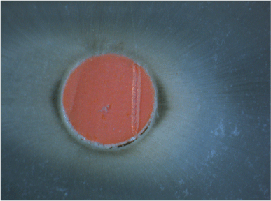
Figure 5: Gap location between the sealer and the root dentin in AH Plus Jet/K3 gutta-percha group.
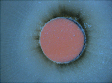
Figure 6: Gaps occurred within the sealer mass in Acroseal/K3 gutta-percha group.
![]()
    n
    Dentin / Sealer
    Core / Sealer
    Within the Sealer
    P values


    Group 1
    270
    224.50
    1029.43
    640.96
    P=0.001


    Group 2
    270
    1431.41
    3.399.23
    1.8667
    P=0.000


    Group 3
    270
    450.90
    197.92
    349.12
    P=0.315


    Group 4
    270
    314.07
    1338.73
    455.12
    P=0.013


    Group 5
    270
    2171.90
    301.97
    323.40
    P=0.000


    Group 6
    270
    248.82
    577.42
    1742.55
    P=0.002


    Group 7
    270
    454.50
    845.90
    306.43
    P=0.018
Table 2: Tukey HSD test results according to the group of regions (mean Μm2). Same letters in the same line indicate statistically similar groups (p>0.05).
Discussion
The results of this study indicated that, leakage inevitably occurs after loss of the coronal seal even when different resin based sealers used in spite of the latest advances in the adhesive-technology as has been previously reported [23]. As well as resin filling materials various physical and biological properties such as; dentin hybridization, polymerization shrinkage, adhesion, adaptability, solubility and antibacterial components, features related with the root canal such as; high configuration factor, smear layer formation due to the instrumentation, regional differences in the quantity, volume, and orientation of the tubules toward the apical portion of the root canal, apical sclerosis, the difficulty in visualization and access to the apical part during primer and material application, restrictions in the flow and distribution of the material may affect resin material's resistance to bacterial penetration by impairing their properties [24,25].
Hybrid Root SEAL sealer was combined both with gutta-percha and Resilon in the present study in order to test if different core materials have any effect on the sealing properties of this relatively new sealer against bacterial penetration. Confirming the results of a previous leakage study [26]. Sixty and six percent of the samples in both groups (gutta-percha and Resilon) showed leakage of E. faecalis within 60-days. This could be because of the similar mechanical properties of gutta-percha/Resilon polymers [27].
Three-D topographical measurements have indicated that Hybrid Root SEAL/Resilon group showed more gaps/voids when compared to the other groups. In a push-out bond strength study, Stiegemeier et al. [28] has pointed numerous voids within the sealer around the core material. According to their failure analysis, the sealer was still intact on the dentinal walls after the test. Onay et al. [29] has also reported that Hybrid Root SEAL showed superior bonding ability to the root dentin with Resilon core material and the failure modes were mostly cohesive within the sealer after the test. In this study, the location of the voids/gaps in Hybrid Root SEAL/Resilon group was mostly between the sealer and the core material. The sealer was continuously observed along the root dentin walls confirming the results of these previous studies [28,29]. According to the results of this study, we can speculate that the leakage might have occurred between the core material and the sealer.
It is obvious from the results of this study that the resistance of RealSeal to bacterial penetration changes according to the core material used (20% with gutta-percha vs 40% with Resilon). Furthermore, median time of leakage is changing greatly in RealSeal groups according to the core material. When gutta-percha was used, median time of leakage was 5-days and when Resilon combined with this sealer, median time of leakage was 41-days. This result confirms the findings of other reports which demonstrated good resistance of this multi-methacrylate sealer with Resilon to bacterial penetration when samples observed for 40-days and 30-days respectively [23,30]. Opposite to our results, unfavorable long term sealing effectiveness of RealSeal / Resilon combination has been reported by Saleh et al. [31] which could be attributable to water uptake [32], bacterial enzymatic degradation and compromised surface integrity with severe surface pitting and erosion of Resilon more than the gutta-percha [33] especially in bacterial leakage set-ups.
RealSeal has been reported to be unable to establish appreciable adhesion to gutta-percha [34]. Steigemeier et al. [28] reported that the failure was 98% between the RealSeal sealer and the root dentin when used with Resilon. However our results showed that RealSeal/gutta-percha group showed the least voids/gaps. Furthermore no significant difference has been found among the location of the gaps or voids. According to Ureyen Kaya et al. [35] the compaction technique can influence the performance of the sealers. In the present study, a matched taper obturation technique has been used. Our results might be different if continuous wave of condensation technique [28], cold lateral compaction technique [35] or System B [29,35] were used.
Although epoxy-resin based AH Plus sealer (AH Plus Jet) was chosen as a reference material in the present study [36], it demonstrated least leak-proof specimens than the others. This could be attributable to the initial setting contraction [37] and debonding of resin from the root canal walls [38] and reduced antibacterial activity of this sealer after setting [39]. In a previous study, Miletic et al. [40] and Ørstavik et al. [37] explained that the leakage of AH Plus by the debonding of the sealer from the root dentin because of shrinkage stresses, initial setting contraction and late expansion of AH Plus sealer that started 4 weeks after setting. In the present study AH Plus Jet/K3 gutta-percha group showed similar gaps/voids with the other groups except Hybrid Root SEAL/Resilon group. The location of the gaps/voids was mostly between the root dentin and the sealer. This result confirms the results of previous findings which indicate that debonding of the resin from the root canal walls may induce leakage.
According to Mutal & Gani [41] the frequency and size of the voids depend on the density of the sealer and increased when the sealers contain calcium hydroxide. In this study, Acroseal/gutta-percha groups showed gaps or voids within the sealer mass. While Acroseal reported to have some antimicrobial activity against E. faecalis [42], 86.7% of this group leaked, confirming the results of Eldeniz and Ørstavik [23]. This could be because of demonstrated solubility, slow ionization due to calcium hydroxide content, polymerization shrinkage and debonding of epoxy-amine resins [23,38].
Diaket sealer also has some inhibitory effect on E. faecalis [43] but only 13.3% of the samples could resist entrance of this bacterium and this could also be referred to solubility and shrinkage of this sealer [44].
In the present study, different sealer/core combinations have been evaluated in terms of leakage and filling quality. In a similar study De Deus et al. [22] have compared the sealing ability of canal filled area on the bacterial leakage of oval-shaped canals and have found no correlation. Present study confirmed De Deus et al. [22] as no correlation has also been found between the results of leakage values and three-D topographical measurements. Van Der Siluis et al. [45] also reported no correlation between fluid filtration leakage test and the percentage of root filling quality. According to them, the percentage of gutta-percha filled area may give a poor image of the root canal filling at the level of the section but the void detected may be cul de sac type and not run from the coronal to the apical therefore not showing fluid transportation. Although the thickness of the diamond saw might have caused extra loss of tooth structure which can be considered as one of the limitations of this study, fifteen sections were obtained from each root in the present study. No correlation between the leakage values and topographical measurements could also be attributable to the cul de sac type gaps/voids as previously mentioned by Van der Sluis et al. [45].
Conclusions
- The present study revealed thNo significant difference was found among the test groups according to their resistance to bacterial leakage.
- The use of Resilon together with the resin based sealer as in this study did not give any advantage over their gutta-percha combinations in preventing bacterial leakage.
- The gaps/voids occurred mostly between the core material and the sealer. All the groups showed similar obturation quality except the MetaSEAL/Resilon group. The mean area of the gaps/ voids occurred at apical sections were smaller than the gaps/ voids occurred at coronal sections. Obturation quality of single-cone technique was found weak at coronal region. In the coronal region use more tapered gutta percha is recommended.
- No correlation exists between the quality of the root filling and bacterial leakage in round-shaped root canals obturated with resin based sealers and a matching tapered single cone technique.
- Among all groups, only the Acroseal group showed gaps or voids within the sealer mass. The use of lateral condensation technique is more appropriate for this sealer.
- In spite of the advanced methacrylate-resin materials, achieving bacteria tight seal is not possible due to the properties of these materials or unfavorable conditions and cavity configuration factor of long, narrow root canals. Further in vivo studies are necessary to evaluate the adhesion between the core materials (Resilon and gutta-percha) and resin based root canal sealers still needs to be improved.
References
- Weller RN, Pashley DH, Tay FR, Loushine RJ. Resistance of a 4-META-containing, methacrylate-based sealer to dislocation in root canals. J Endod. 2008; 34: 833-837.
- Pommel L, About I, Pashley D, Camps J. Apical leakage of four endodontic sealers. J Endod. 2003; 29: 208-210.
- Wu MK, Fan B, Wesselink PR. Diminished leakage along root canals filled with gutta- percha without sealer over time: a laboratory study. Int Endod J. 2000; 33: 121-125.
- Orstavik D, Eriksen HM, Beyer-Olsen EM. Adhesive properties and leakage of root canal sealers in vitro. Int Endod J. 1983; 16: 59-63.
- Kontakiotis EG, Wu MK, Wesselink PR. Effect of sealer thickness on long-term sealing ability: a 2-year follow-up study. Int Endod J. 1997; 30: 307-312.
- Wu MK, Kast'áková A, Wesselink PR. Quality of cold and warm gutta-percha fillings in oval canals in mandibular premolars. Int Endod J. 2001; 34: 485-491.
- El Ayouti A, Achleithner C, Löst C, Weiger R. Homogeneity and adaptation of a new gutta-percha paste to root canal walls. J Endod. 2005; 31: 687-690.
- Zmener O, Pameijer CH, Macri E. Evaluation of the apical seal in root canals prepared with a new rotary system and obturated with a methacrylate based endodontic sealer: an in vitro study. J Endod. 2005; 31: 392-395.
- Gordon M.P.J, Love R.M, Chandler N.P. An evaluation of 06 tapered gutta-percha cones for filling of 06 taper prepared curved root canals. Int Endod J. 2005; 38: 87-96.
- Wu MK, Wesselink PR. Endodontic leakage studies reconsidered. Part I. Methodology, application and relevance. Int Endod J. 1993; 26: 37-43.
- Kersten HW, Moorer WR. Particles and molecules in endodontic leakage. Int Endod J. 1989; 22: 118-124.
- Molander A, Reit C, Dahlén G, Kvist T. Microbiological status of root-filled teeth with apical periodontitis. Int Endod J. 1989; 31: 1-7.
- Siqueira JF Jr, Rôças IN, Souto R, de Uzeda M, Colombo AP. Actinomyces species, streptococci, and Enterococcus faecalis in primary root canal infections. J Endod. 2002; 28: 168-172.
- Eguchi DS, Peters DD, Hollinger JO, Lorton L. A comparison of the area of the canal space occupied by gutta-perchafollowing four gutta-percha obturation techniques using Procosol sealer. J Endod. 1985; 11: 166-175.
- Wu M-K, A.R Özok & Wesselink PR. Sealer distribution in root canals obturated by three techniques. Int Endod J. 2000; 33: 340-345.
- James BL, Brown CE, Legan JJ, Moore BK, Vail MM. An in vitro evaluation of the contents of root canals obturated with gutta percha and AH-26 sealer or Resilon and Epiphany sealer. J Endod. 2007; 33: 1359-1363.
- Gulsahi K, Çehreli Z.C, Onay E, Tasman-Dagli F, Üngör M. Comparison of the area of resin based sealer and voids in roots obturated with resilon and gutta percha. J Endod. 2007; 33: 1338-1341.
- De-Deus G, Murad C, Paciornik S, Reis CM, Coutinho-Filho T. The effect of the canal-filled area on the bacterial leakage of oval-shaped canals. Int Endod J. 2008; 41: 183-190.
- Eldeniz AU, Ørstavik D. A laboratory assessment of coronal bacterial leakage in root canals filled with new and conventional sealers. Int Endod J. 2009; 42: 303-312.
- Bolhuis P, de Gee A, Feilzer A. The influence of fatigue loading on the quality of the cement layer and retention strength of carbon fiber post-resin composite core restorations. Oper Dent. 2005; 30: 220-227.
- Kim YK, Grandini S, Ames JM, Gu LS, Kim SK, Pashley DH, Gutmann JL, Tay FR. Critical review on methacrylate resin-based root canal sealers. J Endod. 2010; 36: 383-399.
- Belli S, Ozcan E, Derinbay O, Eldeniz AU. A comparative evaluation of sealing ability of a new, self etching, dual-curable sealer: Hybrid Root SEAL (MetaSEAL). Oral Surg Oral Med Oral Pathol Radiol Endod. 2008; 106: 45-52.
- Williams C, Loushine RJ, Weller RN, Pashley DH, Tay FR. A comparison of cohesive strength and stiffness of Resilon and gutta-percha. J Endod. 2006; 32: 553-555.
- Stiegemeier D, Baumgartner JC, Ferracane J. Comparison of push-out bond strengths of Resilon with three different sealers. J Endod. 2010; 36: 318-321.
- Onay EO, Ungor M, Ari H, Belli S, Ogus E. Push-out bond strength and SEM evaluation of new polymeric root canal fillings. Oral Surg Oral Med Oral Pathol Radiol Endod. 2009; 107: 879-885.
- Shipper G, Ørstavik D, Teixeira FB, Trope M. An evaluation of microbial leakage in roots filled with a thermoplastic synthetic polymer-based root canal filling material (Resilon). J Endod. 2004; 30: 342-347.
- Saleh IM, Ruyter IE, Haapasalo M, Ørstavik D. Bacterial penetration along different root canal filling materials in the presence or absence of smear layer. Int Endod J. 2008; 41: 32-40.
- Borbely P, Gulabivala K, Knowles JC. Degradation properties and ion release characteristics of Resilon® and phosphate glass/polycaprolactone composites. Int Endod J. 2008; 41: 1093-1100.
- Tay FR, Pashley DH, Loushine RJ, Kuttler S, García-Godoy F, King NM, Ferrari M. Susceptibility of a polycaprolactone-based root canal filling material to degradation. Evidence of biodegradation from a simulated field test. Am J Dent. 2007; 20: 365-369.
- Stoll R, Thull P, Hobeck C, Yüksel S, Jablonski-Momeni A, Roggendorf MJ, Frankenberger R. Adhesion of self-adhesive root canal sealers on gutta-percha and Resilon. J Endod. 2010; 36: 890-893.
- Ureyen Kaya B, Keçeci AD, Orhan H, Belli S. Micropush-out bond strengths of gutta-percha versus thermoplastic synthetic polymer-based systems - an ex vivo study. Int Endod J. 2008; 41: 211-218.
- Brackett MG, Martin R, Sword J, Oxford C, Rueggeberg FA, Tay FR, Pashley DH. Comparison of seal after obturation techniques using a polydimethylsiloxane-based root canal sealer. J Endod. 2006; 32: 1188-1190.
- Orstavik D, Nordahl I, Tibballs JE. Dimensional change following setting of root canal sealer materials. Dent Mater. 2001; 17: 512-519.
- Sevimay S, Kalayci A. Evaluation of apical sealing ability and adaptation to dentine of two resin-based sealers. J Oral Rehabil. 2005; 32: 105-110.
- Pizzo G, Giammanco GM, Cumbo E, Nicolosi G, Gallina G. In vitro antibacterial activity of endodontic sealers. J Dent. 2006; 34: 35-40.
- Miletic I, Anic I, Pezelj-Ribaric S, Jukic S. Leakage of five root canal sealers. Int Endod J. 1999; 32: 415-418.
- Mutal L, Gani O. Presence of pores and vacuoles in set endodontic sealers. Int Endod J. 2005; 38: 690-696.
- Pinheiro CR, Guinesi AS, Pizzolitto AC, Bonetti-Filho I. In vitro antimicrobial activity of Acroseal, Polifil and Epiphany against Enterococcus faecalis. Braz Dent J. 2009; 20: 107-111.
- Eldeniz AU, Erdemir A, Hadimli HH, Belli S, Erganis O. Assessment of antibacterial activity of EndoREZ. Oral Surg Oral Med Oral Pathol Radiol Endod. 2006; 102: 119-126.
- Schäfer E, Zandbiglari T. Solubility of root-canal sealers in water and artificial saliva. Int Endod J. 2003; 36: 660-669.
- Van der Sluis LW, Wu MK, Wesselink PR. An evaluation of the quality of root fillings in mandibular incisors and maxillary and mandibular canines using different methodologies. J Dent. 2005; 33: 683-688.