
Potiguar University – Laureate International Universities, Natal – RN, Brazil
*Corresponding author: Dr. Arcelino Farias-Neto, Health School, Potiguar University, Laureate International Universities, Rua dos Potiguares, 2421, Lagoa Nova, Natal – RN, Brazil,
Received: September 12, 2014; Accepted: November 01, 2014; Published: November 03, 2014
Citation: Farias-Neto A, Dias AHM, de Sousa SA, de Araújo CPD and de S Silva R. An Investigation of the Freeway Space and Facial Proportions in Dentate Subjects. J Dent App. 2014;1(6): 116-118. ISSN:2381-9049
The purpose of this study was to measure the freeway space and facial height of dentate subjects. One hundred and fifty two dentate subjects with ages ranging from 17 to 30 years composed the sample. The freeway space was determined as the difference between rest vertical dimension and occlusal vertical dimension. The Willis gauge was used to measure upper and lower facial heights in resting. Data were collected by two independent observers. The measurements of the two independent observers were submitted to the Intra- Class Correlation test. Differences between upper and lower facial heights were compared with Student’s t test (α = 0.05). The intraclass correlation index (ICC = 0.997, P < .0001) showed excellent reproducibility between the two observers. The freeway space of dentate subjects ranged from 1 to 7 mm. Mean upper facial height (53.7 ± 9.4 mm) and mean lower facial height (54.2 ± 9.9 mm) did not differ significantly. Since most volunteers presented a freeway space ranging from 2 to 4 mm (74%) and the upper facial height seemed to be equal to the lower facial height in resting, these values may be used as initial parameters for the reestablishment of the vertical dimension of occlusion.
Keywords: Facial proportions, physiologic rest position, vertical dimension of occlusion
O objetivo deste estudo foi avaliar o espaço funcional livre a proporção facial em indivíduos dentados. A amostra foi composta por 152 pacientes dentados com idade variando entre 17 e 30 anos. O espaço funcional livre foi calculado como a diferença entre a dimensão vertical de repouso e a dimensão vertical de oclusão. O compasso de Willis foi utilizado para medição da altura dos terços superior e inferior da face em repouso. Os dados foram coletados por dois examinadores independentes e submetidos ao teste de correlação intra-classe. As diferenças entre a altura dos terços superior e inferior da face foram comparadas com o teste T de Student (α = 0.05). O coeficiente de correlação intra-classe mostrou excelente reprodutibilidade (ICC = 0.997, P < .0001). O espaço funcional livre variou de 1 a 7 mm. Não houve diferença significativa entre a altura dos terços superior (53.7 ± 9.4 mm) e inferior da face (54.2 ± 9.9 mm). Visto que a maioria dos pacientes apresentou espaço funcional livre variando de 2 a 4 mm (74%) e a altura dos terços superior e inferior da face em repouso parece ser igual, estes valores poderão ser usados como parâmetros iniciais no restabelecimento da dimensão vertical de oclusão.
Palavras-chave: posição fisiológica de repouso, dimensão vertical de oclusão, proporção facial.
The vertical dimension is defined as the distance between two selected anatomic or marked points, usually one on the tip of the nose and the other upon the chin. The vertical dimension when the mandibular teeth are occluding with the maxillary teeth is defined as the occlusal vertical dimension (OVD). Although controversial, it has been reported that an increased OVD may lead to hyperactivity of the masticatory muscles, elevation in occlusal forces, bruxism and temporomandibular disorders [1]. Nevertheless, changes in OVD certainly affect esthetics and functional activities such as chewing and speech due to their intrinsic relationship with the freeway space and the speaking space. An increased OVD may result from restorative treatment, while a decreased OVD may result from tooth wear or tooth loss [1].
Many methods to reestablish the OVD have been described in the literature. These methods include the use of physiologic rest position, phonetics, esthetics, swallowing, craniometrics, cephalometrics, and electromyography [1,2]. However, there is no single precise scientific method for determining the correct OVD. The physiologic method is widely used due to its simplicity. In that method, the patient must be seated in a comfortable, upright position in the dental chair and completely relaxed to determine the rest vertical dimension (RVD). Then, the OVD is calculated by subtracting from the RVD 2-4 mm related to the freeway space [1]. In the Willis’ method, the RVD is based on facial esthetics and the golden proportion, where the upper facial height (distance between the outer canthus of the eye and corner of the mouth) should be equal to the lower facial height (distance between the lower border of the septum of the nose and lower border of the chin) [1].
The literature suggests that there are some limitations associated with those methods. Johnson et al. [3]. found a wide range for the freeway space (2 to 7 mm), which could lead to inaccurate determination of the OVD when standardized values are used (2-4 mm). When determining the RVD through the physiologic method, different mandibular positions can be obtained at different examination periods for the same individual due to the influence of muscle activity and fatigue [4]. Loss of OVD may be associated with a parallel loss of the vertical dimension when the mandible is at rest, and such a phenomenon would underestimate the freeway space [5]. Attempts to correlate ideal facial proportions with the golden proportion have failed. A three-dimensional study analyzed the faces of professional models and concluded that they did not fit the “golden proportions”, but presented a range of malocclusion and a wide range of cephalometric values [6].
Since more research evidence is required to substantiate the true significance of these concepts in the reestablishment of the OVD, the purpose of the present study was to measure the freeway space and the facial height of dentate subjects. The research hypotheses were that (1) the freeway space of dentate subjects corresponds to the 2-4 mm used in the method of the physiologic rest position, and (2) the upper facial height is equal to the lower facial height in resting, as proposed by the Willis method.
The study was reviewed and approved by the Research Ethics Committee of the institution. The sample was composed by 152 undergraduate students from a Brazilian dental school with ages ranging from 17 to 30 years. Inclusion criteria were natural dentition and bilateral molar support. Subjects presenting tooth wear not compatible with their ages were excluded. For measurement of the freeway space, a small dot was put onto the tip of the nose and chin. Subjects were instructed to sit in the upright position with the head unsupported, swallow and let the jaw relax. RVD (face height with the mandible relaxed and the teeth apart) and OVD (face height with the teeth together) were measured as the distance between these two dots using a divider caliper. The freeway space was determined as the difference between the two measurements. The Willis gauge was used to measure the upper facial height (distance between the outer canthus of the eye and corner of the mouth) and the lower facial height (distance between the lower border of the septum of the nose and lower border of the chin) in resting. All measurements were taken first in the morning to avoid any possibility of muscle fatigue effects on the freeway space [4].
Data were processed with SPSS software (V 17.0 for Windows, SPSS Inc, Chicago, IL, USA). Measurements were accomplished three times for each subject, and the averaged data were used to calculate the distances. Data were collected by two independent observers. Prior to the clinical examinations, 10 volunteers were randomly selected for the calibration process. The measurements of the two independent observers were submitted to the Intra-Class Correlation test. Differences between upper and lower facial height were compared with Student’s t test. Shapiro-Wilk and Levene tests were used to observe normality and variance homogeneity, respectively. Confidence level was set at 95%.
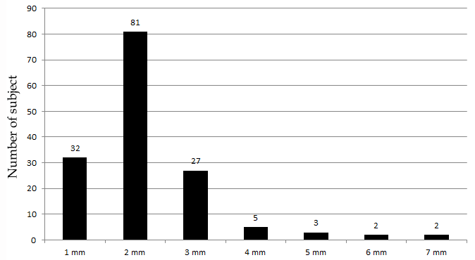
The intraclass correlation index showed excellent reproducibility between the two observers (ICC=0.997, P<.0001). The freeway space of dentate subjects ranged from 1 to 7 mm (Figure 1). Most volunteers presented a freeway space ranging from 2 to 4 mm (74%). Mean upper facial height (53.7 ± 9.4 mm) and mean lower facial height (54.2 ± 9.9 mm) did not differ significantly (p > 0.05).
The results of this study do not support the first research hypothesis that the freeway space of dentate subjects corresponds to the 2-4 mm used in method of the physiologic rest position for reestablishment of the OVD. In the present study, the freeway space of dentate subjects ranged from 1 to 7 mm. These results are in agreement with Johnson et al.3 who also observed a wide range for the freeway space of 72 dentate subjects (2-7 mm). Although most volunteers presented a freeway space ranging from 2 to 4 mm (74%), a large percentage of volunteers presented measurements outside that range (26%). Therefore, it is suggested that the physiologic rest position should be just the first step to determine the OVD.
To ensure adequate freeway space, a combination of physiologic rest position, swallowing, phonetics and esthetics should be employed [1,2]. The swallowing method takes into consideration the individual needs of each patient. It determines the physiologic vertical position from the constantly repeated function of swallowing saliva. During swallowing, the mandible leaves its rest position and rises to the natural OVD. The process of swallowing saliva is recognized as a twenty-four-hour function, and is performed from fifteen hundred to twenty-four hundred times a day. The mandibular pattern of movement is the same for the edentulous infant as it is for the edentulous adultv [7]. In the phonetics method, the patient should be asked to speak while wearing the trial dentures. The teeth do not normally contact during speech, but approach most closely when the ‘S’ sound is made. The separation is known as the smallest speaking space and is usually about 1 mm [1]. Finally, in the esthetics method if the patient’s facial proportions and the contact between upper and lower lips appear to be appropriate when the teeth are occluded, it suggests that the occlusal vertical dimension is correct [1].
The second research hypothesis that the upper facial height is equal to the lower facial height in resting was supported. No significant difference was found between the measurements for the upper (53.7 ± 9.4 mm) and lower (54.2 ± 9.9 mm) facial heights (p > 0.05). These results suggest that the Willis method may be used as an alternative for the physiologic rest position to determine the RVD, preventing that different mandibular positions can be obtained at different examination periods for the same individual due to the influence of muscle activity and fatigue [5]. The results of this study are in contrast to Tina-Olaivar and Olaivar [8] who observed that the upper facial height is longer than the lower facial height by 3 cm when teeth are in occlusion. In another study, only 13% of the dentate subjects examined presented such correlation [9]. These different results may be related to the facial growth pattern (dolichofacial, mesofacial, and brachyfacial) of the samples studied, which was not controlled in any of the studies.
In the present study, measurements of the freeway space were accomplished using a divider caliper and small dots onto the tip of the nose and chin. Sakar et al. [10]. Compared the reliability of two facial measurements, tip of the nose to chin (measured with the divider caliper) and subnasal to chin (measured with the Willis gauge), for determining OVD. The tip of the nose to chin distance presented an improved correlation with the changes in intraoral alterations than subnasal to chin distance. Geerts et al. [2]. compared the accuracy of the Willis gauge with the caliper. Twenty predoctoral students applied both methods 10 times in measuring the RVD and the OVD of a single edentulous patient. The variation of the OVD measurements was significantly smaller for the caliper than for the Willis gauge.
Since many of the proposed techniques for reestablishment of the OVD in dentate subjects have been adapted from complete dentures, further factors must be considered such as remaining tooth structure, space available for the restoration and occlusal variables [11]. The results of the present study were limited to the Brazilian population. More studies are necessary to assess the influence of ethnicity and facial growth pattern on the freeway space and facial proportions.
To summarize, the results of this study suggest the combination of different methods (physiologic, swallowing, phonetics and esthetics) for determining the correct OVD. The method of the physiologic rest position may be used as an initial parameter, since most volunteers presented a freeway space ranging from 2 to 4 mm (74%). Furthermore, the upper facial height seemed to be equal to the lower facial height in resting. It suggests that the Willis method may be used to obtain the RVD, preventing the influence of muscle activity and fatigue inherent to the physiologic method.
Distribution of the freeway space in dentate subjects.
Austin Publishing Group is an emerging open access publisher specialising in Science, Technology and Medicine is dedicated to serve the biomedical community through its initiatives. Austin Publishing Group is an academic publisher with 100+ peer reviewed open access journals in various subjects such as biomedical, Pharma, Life Sciences, Environmental, Engineering and Management. Austin Publishing Group publishes Open Access eBooks providing free access to vast scientific literature.