Case Report
J Dent App. 2015;2(2): 149-152.
Conservative Management of Multiple Odontogentic Keratocyst in a Young Patient with 2 Years Follow Up
Sandeep Ch1, Sindhuri Ve2, Vimala Devi P3 and Nirmala SVSG4*
1Department of Paedodontics and Preventive Dentistry, Former Post Graduate student, Narayana Dental College and Hospital, Nellore, Andhra Pradesh – 524003, India
2Department of Paedodontics and Preventive Dentistry, Post Graduate student, Narayana Dental College and Hospital, Nellore, Andhra Pradesh – 524003, India
3Department of Paedodontics and Preventive Dentistry, Former Post Graduate student, Narayana Dental College and Hospital, Nellore, Andhra Pradesh – 524003, India
4Department of Paedodontics and Preventive Dentistry, Professor, Narayana Dental College and Hospital, Nellore, Andhra Pradesh – 524003, India
*Corresponding author: Nirmala SVSG, Department of Paedodontics and Preventive Dentistry, Professor, Narayana Dental College and Hospital, Nellore, Andhra Pradesh – 524003, India
Received: September 20, 2014; Accepted: January 18, 2015;Published: January 20, 2015
Abstract
Odontogenic keratocyst (OKC) is now designated by World Health Organization (WHO) as keratocystic odontogenic tumor (KCOT). OKC possesses tumor-like characteristics because of its clinical behavior. We report a case of non syndromic multiple OKC in a 14 year old boy with mild mental retardation (M.R). He had swelling since 6 months and pain since 3 days. Intra oral examination revealed obliteration of left labiobuccal vestibule. OPG revealed the presence of seven distinct radiolucent lesions in lower jaw. Histopathologically, it was diagnosed as OKC. Surgical enucleation of the cyst followed by application of Carnoy´s solution. The patient showed uneventful recovery with no recurrence after a follow up of 2 years.
Introduction
Odontogenic keratocyst (OKC) has been the area of interest for many authors since it was first described and termed by Philipsen in 1956 [1]. Earlier in 1926 it was called by other terms such as cholesteatoma by Haur 1926, Kostecka 1929 [2]. The incidence of OKC ranges from infancy to old age with slight male predilection and mandible being the common site of occurrence with specificity in molar ramus area. On the whole the incidence in children in relatively rare when compared to adults, of which sporadic OKC is dominant in incidence than multiple OKC. The management of OKC ranges from the most conservative option like enucleation to aggressive treatment of segmented resection.
We report a case of multiple OKC with 7 individual lesions in mandible of a 14 year old boy who is mentally challenged, which was managed conservatively, with 2 years follow up period.
Case Report
A 14 year old South Indian boy reported to the Department of Pediatric Dentistry with chief complaint of pain and swelling in his left lower back region of the jaw since 3 days. As per the history given by his mother, pain was severe and continuous. On general examination, the child was moderately built and nourished with no signs of pallor, icterus, clubbing, cyanosis and pedal edema.
Extra oral examination revealed gross asymmetry with huge swelling of 7cm X 5cm, extending from left corner of mouth to 1 cm below lobule of ear anteroposteriorly and from 1cm below the ala tragus line to lower border of the mandible superoinferiorly. There was mild increase in cranial circumference and noted with mental retardation. Other major findings like basal nevi, palmar and plantar keratosis, hypertelorism, epidermal cysts of skin are absent. On palpation the swelling was hard with slight increase of temperature, non-compressible and non-fluctuant.
On intra oral examination, left mandibular primary canine was retained with missing mandibular left permanent canine. Left labiobuccal vestibule was obliterated due to swelling of which the mucosa was erythematous. On palpation the swelling is hard and nonfluctuant with foul odor discharge noticed distal to left mandibular second molar.
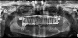
A panoramic radiograph revealed 7 radiolucent lesions with irregular borders were present in the mandible, from mandibular right first permanent molar to left mandibular second permanent molar and extending distally into ramus till 1 cm inferior to sigmoid notch. One of which was associated with mandibular left permanent canine (Figure 1).

Figure 1: OPG showing multiple radiolucencies in the mandible.
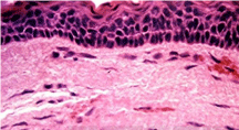
FNAC was done distal to left mandibular second permanent molar which yielded a fowl smelling, thick extract with keratinous plaques along with pus (Figure 2).

Figure 2: Histopathological picture dipicts corrugated parakeratinized stratified squamous epithelium lined with palisaded basal cell layer and connective tissue.
Routine blood investigations revealed elevated erythrocyte sedimentation rate. The biopsy is taken from 3 different areas in relation to Lower left permanent second molar, lower canine and right first permanent molar regions Histopathological examination (Figure 3) of an incisional biopsy specimen revealed that presence of lining epithelium which was very thin and uniform in thickness without evidence of rete ridges, consisted of well defined columnar basal cells in a palisaded arrangement. There was a thin spinous cell layer which shows direct transition from basal cell layer, frequently exhibit intracellular edema. Keratinization was predominantly parakeratotic which was corrugated suggestive of OKC.
Based on clinical, Radiographic and histopathological findings and association with mental retardation, the final diagnosis was given as a multiple OKC in a mentally challenged child.

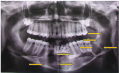
Figure 3: Post operative OPG depicting healing lesions.
Treatment
The boy was taken up for surgery under general anesthesia naso endotracheal intubation was done. Crevicular incision was given from left mandibular second permanent molar to right mandibular first permanent molar and extended along ascending border of ramus distal to former. Fourteen teeth (46 to 37 including 33) were extracted. Thinned out buccal cortical plate was nibbled which exposed the cystic lining. Later, the cystic lining was enucleated followed by curettage and chemical cauterization of the cystic cavity with Carnoy´s solution for 3 minutes. Residual Bone was nibbled and filed followed by smoothening with vulcanite bur. Closure was done with 3.0 vicryl. A small cavity was kept open for regular dressing near mandibular left second permanent molar and packed with sofradyl gauze (chlorhexidine dressing). Patient was followed up for 3 months and dressings were changed for every 2 days, till the cavity healed. Presently, the patient shows no signs of recurrence clinically and radiologically the areas of mandible which once occupied by osteolytic lesions shows radioopacity after 2 years follow up. Currently, we are planning for prosthetic rehabilitation of the child to improve masticatory efficiency and also to prevent supra eruption of the maxillary teeth.
Discussion
After its earlier description as a primordial cyst arising from the remnants of dental lamina mostly in molar angle region of mandible, many studies and publications regarding its variations (clinical, radiological) in presentation and management were put forward. The incidence of OKC although more rare than in adults, they do occur in children. Regezi and Sciubba reported that more commonly multiple OKC´s occur in children as a component of Nevoid Basal Cell Carcinoma Syndrome (NBCCS) [3]. According to Kaban et al. in children with more than one keratocyst, NBCCS must be considered [4]. In our patient, other than mental retardation no other features of NBCCS was present.
In 1960 Gorlin and Goltz [5] first described the spectrum of features associated with NBCC syndrome after which it is also called as Gorlin-Goltz syndrome by McGrath [6]. OKC is also reported to occur in association with other syndromes like Orofacial digital syndrome [7], Ehler-Danlos syndrome [8] and Noonan syndrome [9]. Based on the findings of analysis of clinical features of 312 cases of OKC by, Brannon [10], only 5.8 per cent are patients with multiple keratocysts without other syndrome features, significantly pointing out the lower incidence of non syndromic multiple OKCs. Many such cases are reported in literature by various authors like Auluck [11], Bartake [12], Rudagi [13], Amar [14]. In children they have a wide age range of presentation generally the patients with multiple OKCs with or without NBCCS are generally younger than those with single OKCs. Age of our patient was 14 years and did not present with any of the major clinical findings of any syndrome like nevoid basal cell carcinomas of the skin, bifid ribs, calcification of the falx cerebri, rib anomalies, palmar/plantar keratosis, enlarged head circumference, spina bifida and other features [15] except for multiple odontogenic keratocysts. Unlike other cases our patient is mentally challenged but he is able to point out the pain and swelling and able to respond to our words.
Any swelling in the oral cavity can be primarily evaluated to know its nature by means of FNAC, the aspirate from OKC yields thick fluid which contains keratin plaques and its specific gravity is more than 4mg/ml due to its high protein content. In the present case the aspirate is fowl smelling together with thick keratinous plaques giving an impression of infected OKC.
Routine radiological examinations will help us to know the nature of the lesion especially when it is intraosseous. OKC demonstrates a well defined radiolucent area with smooth and often corticated margins. Large lesions, particularly in the posterior body and ascending ramus of the mandible, may appear multilocular unerupted tooth is involved in the lesion in 25-40% of cases suggesting as dentigerous cyst. Resoption of the roots of adjacent erupted teeth in relation to OKC is less common in comparision to dentigerous and radicular cyst [15].
Main(1970) [16] described radiological varieties of OKC´s like, the one that embraces an adjacent unerupted tooth as envelop mental, those cysts that formed in place of normal tooth was called replacement variety and those in ascending ramus away from teeth was referred as extraneous. Collateral for those OKC´s adjacent to roots of teeth, usually in mandibular premolars.
In our case multiple radio lucencies with well defined sclerotic margins were seen in between mandibular right first premolar and second premolar (first lesion), in relation to root apex of mandibular left first permanent molar, (second lesion), distal to mandibular left second permanent molar root (third lesion), angle and ramus of mandible (4,5,6 lesions), a huge radiolucency measuring around 5 X 3cms is seen associated with crown of impacted mandibular left permanent canine (seventh lesion) giving an impression of dentigerous cyst. None of the adjacent roots of teeth were resorbed. This concludes the impression of multiple cystic lesions along the ength of mandible which was seen more commonly with OKC.
The diagnosis of OKC was mainly based on histopathological features, since the radiological findings although often highly suggestive are not diagnostic. Histologically, OKCs show the presence of a thin band-like parakeratinized or orthokeratinized stratified squamous epithelium, with a prominent basal layer of columnar or cuboidal cells, and an inflammation-free connective tissue wall [15]. Microscopic examination in our case revealed all the features suggestive of OKC.
The relatively early occurrence of multiple OKC may be due to a genetic defect or mutation in the human patched gene [16]. The gene whose mutations cause NBCCS has been mapped to the long arm of chromosome 9q22.1-3-1 [17] and has no apparent heterogeneity. Approximately 50% of cases of NBCCS are associated with allelic losses at this site [17]. The products of this gene may act as a tumor suppressor, as this gene controls growth and development of normal tissues. Reports suggest genetic influences stimulating the formation of OKCs in NBCCS [18]. The occurrence of single or multiple OKCs in the absence of other features of the syndrome represents the least complete form of NBCCS [18,19].
The absence of family history and other features of the syndrome can be due to variation in penetration and expression of different mutations within the same gene or the effects of modifier genes and environmental factors.
The treatment of OKC underwent many series of studies and still it is the most debated topic among the surgeons. Conventional Options include, Enucleation and Curettage, enucleation with chemical cauterization like carnoy´s solution, liquid nitrogen therapy, cryosurgery, Enucleation and Peripheral Ostectomy, Osseous Resection Without (Rim Ostectomy/ Marginal Resection) or With Segmental Resection) Continuity Defect.
The rationale of using carnoy´s solution after enucleation is the same as that of liquid nitrogen that is to remove epithelial remnants and dental lamina in the osseous margin.
The components of Carnoy´s solution are absolute alcohol 6ml, chloroform 3ml, glacial acetic acid 1ml, ferric chloride 1g) . It is a tissue fixative that penetrates bone to a depth of 1.54 mm. Treatment of OKC with peripheral ostectomy, with or without the use of carnoy´s solution, had a significantly lower rate of recurrence. Treatment with enucleation with or without the use of carnoy´s solution was associated with a significantly higher recurrence rate.
Among 20 cysts treated by Stoelinga and Bronkhorst [20] with Enucleation and Carnoy´s, no reported recurrence was found for a period of follow up period of 1-4 years. Similarly, among 40 cases treated by Voorsmit et al. [21], only one case was reported with recurrence for a follow up period of 1-10 years.
Adding Carnoy´s solution to the cyst cavity for 3 minutes after enucleation results in a recurrence rate comparable to that of resection without unnecessarily aggressive surgery.
Simple enucleation was reported to have a recurrence rate of 17% to 56%. Simple enucleation combined with adjunctive therapy, such as the application of Carnoy´s solution or decompression before enucleation, was reported to have recurrence rates of 1% to 8.7% [22].
Conclusion
In any case clinical and radiological follow-up is mandatory for years after surgery because, recurrence may occur even after years. Our patient is on regular follow up and till date showed no signs of recurrence.
References
- P Philipsen HP. Om keratocystedr (Kolesteratomer) and kaeberne. Tandlaegebladet 1956; 60: 963-971.
- Shear M, Speight PM. Cysts of the oral regions. Fourth edition.
- Regezi JA, Sciubba JJ. Oral Pathology clinical pathologic correlations. 5th ed. Saunders
- Kaban LB, Troulis MJ. Pediatric oral and maxillofacial surgery. Saunders; 2004
- Gorlin RJ, Goltz RW. Multiple nevoid basal-cell epithelioma, jaw cysts, and bifid rib syndromes. N Engl J Med 1960; 262: 908-912.
- Mc Grath CJ, Myall RW. Conservative management of recurrent keratocysts in basal-cell naevus syndrome. Aust Dent J 1997; 42: 399-403.
- Lindeboom JA, Kroon FH, De Vires J, Van den Akker HP. Multiple recurrent and De novo Odontogenic keratocysts Associated with oralfacial- digital syndrome. Oral Surg Oral Med Oral Pathol Oral Radiol Endod 2003; 95:458- 462.
- Carr RJ, Green DM. Multiple odontogenic keratocysts in a patient with type II (mitis) Ehler-Danlos syndrome. Br J Oral Maxfacial Surg 1988; 26: 205-214.
- Connor JM, Evans DA, Goose DH. Multiple odontogenic keratocysts in a case of the Noonan syndrome. Br J Oral Surg 1982; 20: 213-216.
- Brannon RB. The odontogenic keratocyst. A clinicopathologic study of 312 cases.Part I. Clinical features. Oral Surg Oral Med Oral Pathol 1976; 42: 54-72.
- Auluck A, Suhas S, Pai KM. Multiple odontogenic keratocysts: report of a case. J Can Dent Assoc 2006; 72: 651-656.
- Bartake AR, Shreekanth NG, Prabhu S, Gopalkrishnan S. Non- Syndromic Recurrent Multiple Odontogenic Keratocysts: A Case Report. J Dent (Tehran) 2011; 8: 96-100.
- Rudagi BM, Kharkar VR, Kini Y. Multiple odontogenic keratocysts with Diverse histologic features in a non- syndromic patient. Pravara Med Rev 2010; 5: 35-37.
- Sholapurkar AA, Varun RM, Pai KM, Geetha V. Non-syndromic multiple odontogenic keratocysts: report of case. Rev Clín Pesq Odontol 2008; 4: 193-199.
- Neville BW, Damm DD, Allen CM, Bouquot JE. Oral and maxillofacial pathology. 2nd ed. Philadelphia: Saunders; 2002.
- Lench NJ, Telford EA, High AS, Markham AF, Wicking C, et al. Characterisation of human patched germ line mutations in naevoid basal cell carcinoma syndrome. Hum Genet 1997; 100: 497-502.
- Lo Muzio L, Nocini P, Bucci P, Pannone G, Consolo U, Procaccini M. Early diagnosis of nevoid basal cell carcinoma syndrome. J Am Dent Assoc 1999; 130: 669-674.
- el Murtadi A, Grehan D, Toner M, McCartan BE. Proliferating cell nuclear antigen staining in syndrome and nonsyndrome odontogenic keratocysts. Oral Surg Oral Med Oral Pathol Oral Radiol Endod 1996; 81: 217-220.
- Shear M. Odontogenic keratocyst: natural history and immunohisto¬chemistry. Oral Maxillofac Surg Clin N Am 2003; 15: 377-382.
- Stoelinga PJW, Bronkhorst FB. The incidence, multiple presentation and recurrence of aggressive cysts of the jaws. J Craniomaxillofac Surg 1988; 16: 184-195.
- Voorsmit RA, Stollinga PT, Van Hallst VJ. The management of keratocysts. J Maxillofac Surg 1981; 9: 228-236.
- Blanas N. Systematic review of the treatment and prognosis of the odontogenic keratocyst. Oral Surg Oral Med Oral Pathol Oral Radiol Endod 2000; 90: 553-558.