
Case Report
J Dent App. 2015;2(5): 229-231.
Intraoral Verrucous Hemangioma: A Case Report
Lele GS1*, Salunkhe BB2 and Lele SM3
1Professor and Head, Department of Pediatric and Preventive Dentistry, Sinhgad Dental College and Hospital, India
2Postgraduate student, Department of Pediatric and Preventive Dentistry, Sinhgad Dental College and Hospital, India
3Professor, College of Dentistry, King Faisal University, Saudi Arabia
*Corresponding author: Lele GS, Department of Pediatric and Preventive Dentistry, Sinhgad Dental College and Hospital, Vadgaon Budruk, Pune 411041,India
Received: January 15, 2015; Accepted: March 27, 2015; Published: March 30, 2015
Abstract
Verrucous hemangioma is a lesion with a very rare occurrence in the oral cavity. It is commonly found on the extremities. This case report describes a rare case of verrucous hemangioma present intraorally in a pediatric patient.
A 13 year old male patient reported with a complaint of lump in lower left back tooth region with associated difficulty in swallowing. On clinical examination, a proliferative growth, bluish black in colour was seen distal to mandibular left second permanent molar. Papules were observed on the soft palate extending into pharynx. Radiographic investigations and incisional biopsy was performed. Radiographic and histopathological evaluation followed by immunohistochemistry analysis helped to confirm the diagnosis as verrucous hemangioma.
Keywords: Verrucous hemangioma; Intraoral; Pediatric patient; Retromolar; Papules; Pharynx; Angiokeratoma
Introduction
Verrucous hemangioma was first described by Halter in 1937. Loria et al defined this entity in 1958, and in 1967, Imperial and Helwig introduced the term ‘verrucous hemangioma’ and defined it as a congenital vascular malformation comprising a capillary or cavernous hemangioma in the dermis and subcutaneous tissue associated with reactive epidermal acanthosis, papillomatosis and hyperkeratosis [1]. It has been reported in literature with a variety of names such as, hemangioma unilateralis neviforme, unilateral verrucous hemangioma, angiokeratoma circumscriptum neviforme, nevus vascularis unius lateralis, keratotic hemangioma, nevus angiokeratoticus, nevus keratoangiomatosus and papulous angiokeratoma.
Case Report
A 13 year-old male patient reported to the department of Paediatric and Preventive Dentistry at Sinhgad Dental College and Hospital, Pune, with a chief complaint of a lump present in lower left back tooth region since 3 days. It caused pain during swallowing and had gradually increased to the present size. The patient was of normal built and stature, with a non-contributory family and medical history. He had no past dental history. On clinical examination, a proliferative growth was seen extending from distal aspect of 37 involving the pterygomandibular and retromolar areas, and was approximately 3 centimeters by 2 centimeters in size. The lesion appeared to be reddish pink in colour, interspersed with bluish black areas grouped together. Surface of the growth was irregular. Papules were seen on soft palate extending upto the pharynx which caused difficulty in swallowing and discomfort (Figure 1). On palpation, the lesion was soft and compressible. Left submandibular lymph nodes were palpable, nontender, enlarged, mobile, soft and not fixed to underlying structures. Intraoral radiographic investigations did not show any significant findings. The panoramic radiograph (Figure 2) showed no periapical or alveolar pathology. Incisional biopsy was carried out under local anesthesia and subjected to histopathological evaluation and immunohistochemistry. On dermatologic examination, no lesions were found anywhere on the skin.
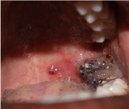
Figure 1: Intraoral appearance of proliferative growth distal to 37.
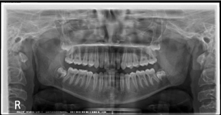
Figure 2: Panoramic radiograph showing no pathology.
Histopathological findings showed stratified squamous hyperkeratinized epithelium with hyperplasia and papillary projections above the surface. Elongated rete ridges were noted. Fibro cellular connective tissue with numerous small and large vascular spaces lined by endothelial cells and filled with RBC’s was seen. Dilated blood filled spaces were present in the sub-epithelial connective tissue and in the deeper connective tissue proliferating around the muscle. Connective tissue seemed to be infiltrated with chronic inflammatory cells and mast cells (Figures 3a and 3b). Immuno histochemistry analysis was finally performed to confirm the diagnosis and it showed strong positivity with CD34 marker in the deeper layers too (Figure 4). Thus the diagnosis of an intraalveolar verrucous hemangioma was established.
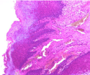
    Figure 3 a:  Hematoxylin and Eosin staining under 10 X magnification – Image
of H&E stained section showing hyperkeratosis, elongated rete ridges and
numerous dilated capillaries.
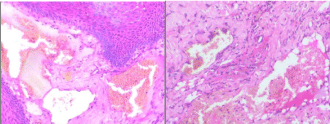
    Figure 3 b:  Hematoxylin and Eosin staining under 40X magnification : Image
of H&E stained section showing deeper layers with numerous dilated blood
filled spaces.
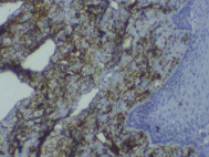
    Figure 4:  Immunohistochemistry analysis under 40X magnification:
Photomicrograph showing strong positivity with CD34 marker in the deeper
areas too.
Discussion
Verrucous hemangioma is seen at birth or during infancy, but it may occur in adulthood too. It can start as lesions resembling portwine stains which may later become soft bluish- red vascular swellings that tend to grow in size and become verrucous [2]. Helmig et al. [3] observed that verrucous hemangioma presents usually as a warty blue-black lesion in the lower extremities of children. The lesions tends to be unilateral, well-defined, discrete, grouped, bluish-red, soft, and compressible; they vary between 4 millimeters to 7 centimeters in diameter and small satellite lesions are often present. Clinically, they may resemble angiokeratoma, lymphangioma circumscriptum, verrucous epidermal nevus, verrucous cancer or even malignant melanoma [4].
Histologically, verrucous hemangioma presents an epidermis with irregular acanthosis and hyperkeratosis. The abnormal vessels are located in the dermis and hypodermis, and extend along the vertical vascular channels with almost no involvement of the reticular dermis. The vessels are round, with thick walls and a multi lamellar basement membrane. The dilated vessels of the papillary dermis often contain blood, are thin-walled, and have a vertical orientation, whereas the deeper vessels may contain blood or be empty [5]. Secondary infection and bleeding is a frequent complication and this could result in reactive papillomatosis and hyperkeratosis, and thus, the older lesions acquire a verrucous or warty surface. Unlike other angiomatous nevi, they do not involute spontaneously [6]. A biopsy of sufficient depth is required in this type of lesion, as a superficial tissue sample may lead to an erroneous initial diagnosis, with the consequent inappropriate treatment and relapse [7].
The differential diagnosis includes Cobb syndrome, angioma serpiginosum, lymphangioma circumscriptum, cutaneous keratotic hemangioma, blue rubber bleb nevus, papillomas, and tumors including melanoma [8]. However, the main differential diagnosis must be made with angiokeratoma circumscriptum. Verrucous hemangioma is usually a solitary lesion that varies from 1 to 7 centimeters in diameter and is often surrounded by smaller satellite lesions; angiokeratoma circumscriptum, on the other hand, is typically formed of punctate lesions that vary between 1 and 5 mm in diameter and that occasionally coalesce to form plaques several centimeters across [9]. The main histological difference is that verrucous hemangioma extends into the hypodermis whereas angiokeratoma circumscriptum is limited to the superficial dermis [10]. Immunohistochemically, the endothelium shows focal positivity for type 1 glucose transporter and low-level reactivity for mindbomb homolog 1. In contrast, it does not stain with D2-40 [11]. Angiokeratoma circumscriptum can be treated with the usual physical methods such as electrocoagulation, cryotherapy, and argon laser, verrucous hemangioma requires wide excision to avoid possible recurrence [12]. The patient was referred to ENT department for further management.
References
- Imperial R, Helwig EB. Verrucous hemangioma. A clinicopathologic study of 21 cases. Arch Dermatol 1967; 96: 247-253.
- Popadic M. Evolution of verrucous hemangioma. Indian J Dermatol Venereol Leprol 2012; 78: 520.
- Nandaprasad S, Sharada P, Vidya M, Karkera B, Hemanth M, et al. Hemangioma-A review. The Internet J Hematol 2008; 6.
- Yasar A, Ermertcan AT, Bila C, Bila DB, Temiz P. et al. Verrucous hemangioma. Indian J Dermatol Venereol Leprol 2009; 75: 528-530.
- Garrido-Ríos AA, Sánchez-Velicia L, Marino-Harrison JM, Torrero-Antón MV, Miranda-Romero A. A Histopathologic and Imaging Study of Verrucous Hemangioma. Actas Dermosifiliogr 2008; 99: 723-726.
- Wang G, Chunying L, Gao T. Verrucous hemangioma. Int J Dermatol 2004; 43: 745-746.
- Achar A, Biswas SK, Maity AK, Naskar B. Verrucous hemangiomatreated with electrocautory. Indian J Dermatol 2009; 54: 51-52.
- Jain VK, Aggarwal K, Jain S. Linear verrucous hemangioma on the leg. Indian J Dermatol Venereol Leprol 2008; 74: 656-658.
- Garrido-Rios AA, Sanchez-Velicia L, Marini-Harrison JM, Torrero-Anton MV, Miranda-Romero A. Verrucous hemangioma: Histopathological and radiological study. Proceedings Dermosifiliogr 2008; 99: 723-726.
- Pavithra S, Mallya H, Kini H, Pai GS. Verrucous hemangioma or angiokeratoma? Missed diagnosis. Indian J Dermatol 2011; 56: 599-600.
- Tennant LB, Mulliken JB, Perez-Atayde AR, Kozakewich HP. Verrucous hemangioma revisited. Pediatric Dermatol 2006; 23: 208-215.
- Bhat S, Pavithra S, Mallya H, Pai G. Verrucous hemangioma: an optimized surgical approach. J Cutan Aesthet Surg 2010; 3: 170-173.