
Abstract
The pathological changes of the salivary glands include developmental anomalies, sialadenitis, sialolithiasis, mucous retention and neoplastic lesions. Bacterial sialadenitis is an important head and neck infection. Sialolithiasis is the obstruction of the excretory system of a gland by calcified elements within it. The objective of this paper is to present a case of submandibular sialadenectomy in a patient with both infectious sialadenitis and sialolithiasis. A 54-year-old female presented with a history of 10 years of swelling, pain, and drainage of pus in the sublingual region, leading to hospital admissions. The clinical examination showed a symptomatic swelling of the right submandibular region, with purulent secretions from the submandibular duct. Radiographic examination demonstrated a calculus in the floor of the mouth. Ultrasonography showed glandular enlargement with heterogeneous texture. The patient was treated with submandibular sialadenectomy using the transcervical approach. A calculus was removed from the submandibular duct. Histopathological examination showed sialadenitis and sialolithiasis with the formation of smaller stones within the gland. The patient recovered well and remained asymptomatic at the time of her one-year follow-up.
Keywords: Salivary glands; Submandibular gland; Sialadenitis, Sialolithiasis
Introduction
The submandibular glands are subject to several pathologies that require its excision. The most common problems that affect these glands are sialadenitis and sialolithiasis [1,2]. The excision of the submandibular gland has been indicated in cases of sialoliths, sialadenitis and tumors [3].
Salivary gland infections are of bacterial or viral etiology, being observed at all ages [4,5]; their presentation can be acute or chronic, while acute suppurative sialadenitis presents as rapid-onset pain and swelling [6].
Sialolithiasis is characterized by the development of calcified structures in the salivary glands, usually in adults. Sialoliths are accompanied by swelling and pain when a salivary stimulus occurs [5].
For calculi located near the ostium, catheterization facilitates their removal [7]. For sialoliths located in the anterior half of the duct, removal is recommended by intra-oral access. Those located in the posterior portion of the duct or intraglandular sialoliths sometimes require the removal of the gland [8].
The objective of this paper is to present a case of submandibular sialadenectomy in the simultaneous occurrence of infectious sialadenitis and sialolithiasis.
Case Presentation
A 54–year-old woman was referred with complaints of pain and swelling in the right submandibular region and intra-oral drainage of pus. She reported a history of 10 years of symptom evolution, with recurrent episodes and hospital admissions. Her medical history was uneventful.
On examination, facial asymmetry was observed with symptomatic welling of the right submandibular gland, and gland milking resulted in purulent discharge (Figure 1). The mandibular occlusal radiograph showed an image suggestive of a calculus (Figure 2).
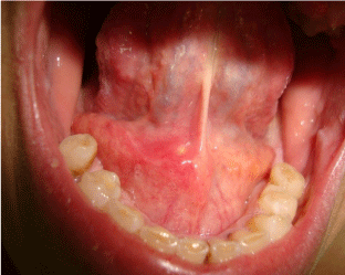
    Figure 1:  Intraoral aspect showing reddish swelling near the exit of the
Submandibular duct.
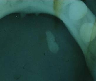
    Figure 2:  Irregular radiopaque image observed in the mandibular occlusal
radiograph duct indicative of a calculus.
The ultrasound test showed the right submandibular gland with increased dimensions and a heterogeneous texture, indicating sialadenitis. The diagnosis of infectious sialadenitis associated with sialolithiasis was made. Amoxicillin and metronidazole were prescribed to control the infection. Removal of the affected submandibular gland was proposed, considering the diagnosis, the time of symptom evolution and previous treatments.
The surgery was performed under general anesthesia, using the transcervical approach. The procedure comprised accessing the right gland (Figure 3), submandibular duct ligature, and excision of the gland (Figure4). A large calculus was removed from the submandibular duct through a small incision in the mouth floor.

    Figure 3:  Intra operative aspect of the dissection of the right Submandibular
gland

    Figure 4:  Individualization of the duct aiming its ligation prior to excision of
the submandibular gland.
Microscopic findings showed foci of intense mononuclear inflammatory infiltrate permeating the ducts and secretory units (Figure 5 and 6). Within a duct there was a nodule consisting of concentric laminations of acellular eosinophilic material (Figure 7). The diagnosis was chronic sialadenitis and sialolithiasis.

    Figure 5:  General view of submandibular gland showing dilated excretory
ducts (H & E, x 100).
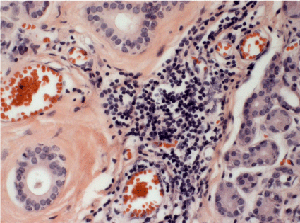
    Figure 6:  Interlobular area of the gland exhibiting an intense mononuclear
infiltrate (H & E, x 400).
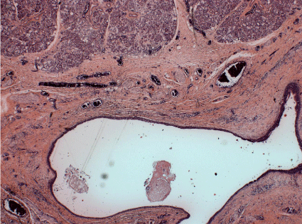
    Figure 7:  A detail of an excretory duct showing a partially mineralized
material (sialolith) in its lumen (H & E, x 400).
After seven days there was mild edema in the right submandibular region. Three months postoperatively the patient was asymptomatic, with satisfactory scarring of the incisions. One year postoperatively, the patient had no complaints and was satisfied with the treatment performed (Figure 8).
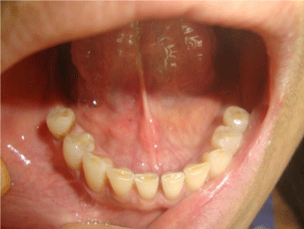
Figure 8: Intraoral aspect showing characteristics of normality in the mouth floor.
Discussion
A case of submandibular sialadenectomy in a patient with both infectious sialadenitis and sialolithiasis is reported. These diagnoses are usually confirmed with clinical and radiological examination. Other imaging resources can provide additional information [6,9].
Diagnosis of salivary disorders begins with a careful examination [10]. Swelling and stiffness are frequent signs of infectious sialadenitis [11]. In the present case, facial asymmetry, symptomatic swelling and the expression of pus led to the diagnosis of infectious submandibular sialadenitis.
Acute suppurative sialadenitis has a polymicrobial etiology. The bacteria may be aerobic or facultative, anaerobic, or mixed aerobic and anaerobic [12]. Penicillinase-resistant penicillin or a firstgeneration cephalosporin is adequate for this infection [12].
Additionally, a calculus was detected on palpation. This allowed its intraoral removal [7]. An image of a calculus was detected by mandibular occlusal radiograph- a suitable imaging modality for stones in the anterior portion of the duct. Ultrasound test showed increased dimensions and heterogeneous texture of the gland, indicating sialadenitis.
The ultrasound test is also helpful in the assessment of Sialolithiasis [9]. Usually, in sialolithiasis only one calculus is found [13]. Our finding of one calculus in the duct and the other inside the gland configures a less common situation.
The conservative treatment of salivary calculi includes extracorporeal shock-wave lithotripsy, fluoroscopically guided basket retrieval or intraoral stone removal under general anesthesia, used either alone or in combination [14].
The main reasons for the surgical excision of the gland were: long-standing sialoliths which usually produce irreversible functional damage [15] and the recurrent infections of the gland [5]. The advantages of the chosen treatment are complete resolution of pathological changes and remission of symptoms, and the disadvantages are the possible risks, not seen, of nerve damage [3,16].
References
- Rallis G, Mourouzis C, Zachariades N. A study of 55 submandibular salivary gland excisions. Gen Dent 2004; 52: 420-423.
- Torroni AA, Mustazza MC, Bartoli DD, Iannetti GG. Transcervical submandibular sialoadenectomy. J Craniofac Surg. 2007; 18: 613-621.
- Preuss SF, Klussmann JP, Wittekindt C, Drebber U, Beutner D, Guntinas-Lichius O. Submandibular gland excision: 15 years of experience. J Oral Maxillofac Surg. 2007; 65: 953-957.
- Turner MD, Glickman R. Case presentations of salivary gland infections. Oral Maxillofac Surg Clin North Am. 2009; 21: 359-362.
- Delli K, Spijkervet FK, Vissink A. Salivary gland diseases: infections, sialolithiasis and mucoceles. Monogr Oral Sci. 2014; 24: 135-148.
- Wilson KF, Meier JD, Ward PD. Salivary gland disorders. Am Fam Physician. 2014; 89: 882-888.
- Park JS, Sohn JH, Kim JK. Factors influencing intraoral removal of submandibular calculi. Otolaryngol Head Neck Surg. 2006; 135: 704-709.
- Komatsuzaki Y, Ochi K, Sugiura N, Hyodo M, Okamoto A: Video-assisted submandibular sialadenectomy using an ultrasonic scalpel. Auris Nasus Larynx. 2003; 30: S75-78.
- Alyas F, Lewis K, Williams M, Moody AB, Wong KT, Ahuja AT, et al. Diseases of the submandibular gland as demonstrated using high resolution ultrasound. Br J Radiol. 2005; 78: 362-369.
- Arduino PG, Carrozzo M, Pentenero M, Bertolusso G, Gandolfo S. Non-neoplastic salivary gland diseases. Minerva Stomatol. 2006; 55: 249-270.
- Duker J. Sialadenitis of the left submandibular gland/siallolith. Quintessence Int. 2005; 36: 747-748.
- Brook I. Aerobic and anaerobic microbiology of suppurative sialadenitis. J Med Microbiol. 2002; 51: 526-529.
- Choi J, Kim IK, Oh NS. Multiple sialolitis in sublingual gland: report of a case. Int J Oral Maxillofac Surg. 2002; 31: 562-563.
- McGurk M, Escudier MP, Brown JE: Modern management of salivary calculi. Br J Surg.2005; 92: 107-112.
- El Gehani R, Krishnan B. Shehoumi MI: Submandibular giant sialoliths: report of two cases and review of the literature. Ear Nose Throat J. 2010; 89: E1-4.
- Hernando M, Echarri RM, Taha M, Martin-Fragueiro L, Hernando A, Mayor GP. Surgical complications of submandibular gland excision. Acta Otorrinolaringol Esp. 2012; 63: 42-46.