Case Report
J Dent & Oral Disord. 2016; 2(8): 1043.
Minimally Invasive Technique to Mask White Spot Lesion with Resin Infiltration (Icon®, DMG): Case Report
Fonseca NMAH¹, Públio JC¹, Hernandes DKL¹, Barros TEP¹, Borelli Neto L¹ and Alves N2,3*
¹Guarulhos Faculty of Sciences, Guarulhos, Brazil
²CIMA Research Group, Temuco, Chile
³Department of Dentistry, Universidad de La Frontera, Chile
*Corresponding author: Nilton Alves, Department of Dentistry, Universidad de La Frontera, Temuco, Chile
Received: October 11, 2016; Accepted: November 14, 2016; Published: November 15, 2016
Abstract
The case report describes a minimally invasive technique to mask white spots lesion with resin infiltration Icon, DMG. The etching procedure on the white spot lesion exposes the microporosities, permitting the penetration of lowviscosity light-curing resins by capillary force. When the lesions are infiltrated, the microporosities are occluded and the whitish appearance disappears. The use of this technique is an alternative to restorative treatment and the micro abrasion.
Keywords: White spot lesions; Caries infiltration; Aesthetics
Introduction
White Spot Lesions (WSP) are detected frequently and can be problematic for the patients with important esthetic concerns [1]. WSP are the earliest clinically evident manifestation of the caries process, exhibiting subsurface porosity caused by an imbalance between the biological dynamic processes of demineralisation and remineralisation [2,3].
These lesions develop as a result of prolonged plaque due to inadequate oral hygiene. The insertion of fixed orthodontic appliances creates stagnation areas for plaque and makes tooth cleaning more difficult; therefore, for success the patient must be more careful with oral hygiene and use preventive strategies involving oral health promotion.
White spot prevalence of 50% [4], 60% [5] or even 97% [6] after bonded or banded orthodontic treatments has been reported and can appear on any tooth surface, however, most frequently occurs on the vestibular surfaces of anterior teeth causing esthetic problems [7-9].
Several techniques have been proposed to mask the appearance of white spot lesions and some authors demonstrated that infiltration treatment was capable to cosmetically camouflage enamel caries lesions [10,11].Treatment for white spot lesions comprises restorative procedures, improvement of remineralization using a complex of casein phosphopeptides and amorphous calcium phosphate (CCPACP) or flouride-containing products, microabrasion, argon-laser irradiation [12].
The aim of this study was to report a case of treatment using the technique to mask white spots lesion with resin infiltration Icon® (DMG), after orthodontic treatment.
Case Presentation
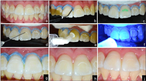
An 18-year-old female patient came to Guarulhos Faculty of Sciences- Brazil, to correct the white lesions on her upper and lower teeth. Patient’s history included orthodontic treatment for 2 years, but the orthodontic professional decided to stop orthodontic treatment due to that the patient presents inadequate oral hygiene. The oral examination, that was performed after dental prophylaxis, revealed cavities on the cervical third of 31 and 32 and carious white spot lesion on the upper and lower anterior teeth (Figure 1a). Following the manufacturer’s instructions, Icon® (DMG, Hamburg, Germany) was applied. Initially, a rubber dam was applied to protect soft tissue (Figure 1b), followed by an application of Icon Etch (15% hydrochloric acid gel) for 2 min on the white spot lesion (Figure 1c). The etching gel was washed for 30s using a water spray. The lesion is dried by applying ethanol (Icon-Dry) for 30s followed by air drying (Figure 1d). Infiltrant was applied for 3 min (Figure 1e). The Icon® (DMG, Hamburg, Germany) application was repeated for 1 minute to minimize enamel porosity. Both applications were light cured for 40s (Figure 1f). Figure 1g shows the results obtained immediately after the Icon application. The procedure was repeated in the other incisor. Figures 1h and 1i show the aspect before Icon application and the immediate post-operative, respectively. We observed that, despite the appearance of the enamel was not 100% reestablished, aesthetic appearance improved significantly.

    Figure 1:  Preoperative condition of the patient (a); Rubber dam is applied to
protect soft tissue (b); Application of Icon Etch (15% hydrochloric acid gel) on
the white spot lesion (c); The lesion is dried by applying ethanol (Icon-Dry) for
30 s (d); Application of infiltrant (e); Both applications were light cured for 40
s (f); Immediately after the Icon application (g); Before Icon application (h);
Immediate post-operative (i).
Discussion
Development of WSL is a challenging problem during the course of orthodontic treatment [13]. Even with rigorous oral hygiene WSL may arise, leading to permanent aesthetic damage [14]. The loss of minerals in WSL creates porosities that change the refractive index of the enamel [15]. Paris et al. [16] suggests that in case of WSL post-orthodontic treatment, infiltration should be done as quickly as possible. Resin infiltration is a new micro invasive approach to arrest the progress of proximal initial caries lesions [17]. The advantages of resin infiltration are that the procedure does not require anesthesia, is quick, painless and returns the aesthetic due to that the white spots lose their caulky appearance.
The technique using Icon® DMG advocates the etching procedure to remove mineral of the surface layer, less than 30mm demineralized enamel [17]. The purpose of the etching procedure is to expose the lesion porosities there for elow-viscosity light-curing resin can infiltrate, filling microporosities of the white spot lesion and replacing the initial appearance of the enamel. A positive side effect of resin infiltration is that enamel lesions lose their whitish appearance when their microporosities are filled with the resin and look similar to sound enamel [1]. The infiltrant (Icon®, DMG) can be used for both the vestibular and interproximal noncavitated lesions [18].
Resin infiltration presents an argument to the therapeutic spectrum of orthodontists, as well as pediatric or general dentists, in that enamel are as affected by post-orthodontic WSLs can be restored to their original appearance [19].
Minimal invasive dentistry by resin infiltration technique seems to provide a good solution in treating early enamel lesions [18]. The ultimate goal of treating discoloration of teeth is to get an acceptable aesthetic result in the most conservative way possible [20]. Sandoval et al. [19] claim that the aesthetic appearance of WSL post-orthodontic is improved by resin infiltration, being the best results achieved with infiltration of mild to moderate WSL directly or closely following the appointment of debonding. Kim et al. [21] analyzed the effect of resin infiltration in 20 teeth with a developmental defect of enamel and 18 teeth with post orthodontic descalcification, and observed that 25% of teeth were classified as completely masked, while 35% were partially masked and were 40% unchanged. In the present study, we observed a significant improvement in restoring enamel and aesthetics of the teeth.
A limitation of this technique is the need to follow accurate diagnosis criteria to distinguish between the developmental and non-developmental opacities, because the resin infiltration shows limited effects in cases of developmental defects; furthermore it is a radiolucent material, which may be a concern to some dentists. These factors determine the success of treatment [22]. It is also important to consider that the depth of resin infiltration is 60, therefore the best treatment must be assessed for the elimination of white spots given deeper lesions could be detected even after treatment with resin infiltration [23].
Gugnani et al. [18] state that patient’s motivation would probably play a major role in the success of any minimally invasive technique. In our study, the patient will return for new investments and, after the restorative treatment, she will be able to return to the orthodontic treatment.
We agree with Gugnani et al. [18] and Kim et al. [21] in their claims that further investigations with longer periods of follow up are necessary, in order to confirm the efficiency of this treatment modality and encourage the clinicians to use in their dental practice.
Conclusion
The treatment of white spot lesions with infiltrating procedure using Icon (DMG, Hamburg, Germany) is easy, quick, and painless and changes the appearance of the white spot lesion thereby giving an excellent cosmetic result.
References
- Jeong-Hye Son, Bock Hur, Hyeon-Cheol Kim, Jeong-Kil Park. Management of white spots: resin infiltration technique and microabrasion. JKACD. 2011; 36: 66-71.
- Featherstone JD. The science and practice of caries prevention. J Am Dent Assoc. 2000; 131: 887-899.
- Kidd EA, Fejerskov O. What constitutes dental caries? Histopathology of carious enamel and dentin related to the action of cariogenic biofilms. J Dent Res. 2004; 83: 35-38.
- Gorelick L, Geiger AM, Gwinnett AJ. Incidence of white spot formation after bonding and banding. Am J Orthod. 1982; 81: 93-98.
- Hadler-Olsen S, Sandvik K, El-Agroudi MA, Øgaard B. The incidence of caries and white spot lesions in orthodontically treated adolescents with a comprehensive caries prophylactic regimen - a prospective study. Eur J Orthod. 2011; 34: 633-639.
- Boersma JG, van der Veen MH, Lagerweij MD, Bokhout B, Prahl-Andersen B. Caries prevalence measured with QLF after treatment with fixed orthodontic appliances: influencing factors. Caries Res. 2005; 39: 41-47.
- Tufekci E, Dixon JS, Gunsolley JC, Lindauer SJ. Prevalence of white spot lesions during orthodontic treatment with fixed appliances. Angle Orthod. 2011; 81: 206-210.
- Torres CRG, Borges AB, Torres LMS, Gomes IS, de Oliveira RS. Effect of caries infiltration technique and fluoride therapy on the colour masking of white spot lesions. J Dent. 2011, 39: 202-207.
- O'Reilly MT, De Jesús Viñas J, Hatch JP. Effectiveness of a sealant compared with no sealant in preventing enamel demineralization in patients with fixed orthodontic appliances: A prospective clinical trial. Am J Orthod Dentofacial Orthop. 2013; 143: 837-844.
- Paris S, Meyer-Lueckel H. Masking of labial enamel white spot lesions by resin infiltration—a clinical report. Quintessence Int. 2009; 40: 713-718.
- Kim S, Kim EY, Jeong TS, Kim JW. The evaluation of resin infiltration for masking labial enamel white spot lesions. Int J Paediatr Dent. 2011; 21: 241-248.
- Cha SW, Yoon TC, Park SH, Lee CY, Kum KY. Quantitative analysis of mineral change in the initial car10us lesion using confocal laser scanning microscopy. J Korean AcadConservDent. 2001; 26: 1-8.
- Willmot D. White spot lesions after orthodontic treatment. Seminar in Orthodontic. 2008; 14: 209-219.
- Hammad SM, El Banna M, El Zayat I, Mohsen MA. Effect of resin infiltrationon white spot lesions after debonding orthodontic brackets. Am J Dent. 2012; 25: 3-8.
- Featherstone JD. Prevention and reversal of dental caries: Role of low level fluoride. Community Dent Oral Epidemiol. 1999; 27: 31-40.
- Paris S, Meyer-Lueckel H, Kielbassa AM. Resin infiltration of natural caries lesions. J Dent Res. 2007; 86: 662-666.
- Meyer-Lueckel H, Paris S, Kielbassa AM. Surface layer erosion of natural caries lesions with phosphoric and hydrochloric acid gels in preparation for resin infiltration. Caries Res. 2007; 41: 223-230.
- Gugnani N, Pandit IK, Gupta M, Josan R. Caries infiltration of noncavitedwhite spot lesions: a novel approach for immediate esthetic improvement. Contemp ClinDent. 2012; 3: 199-202.
- Sandoval P, Vogel R, Henríquez D, Knösel M. Management o post-orthodontic white-spot-lesions: clinical handling of the resin infiltration technique (Icon®, DMG). Int J Odontostomatol. 2016; 10: 29-33.
- Muñoz MA, Arana-Gordillo LA, Gomes GM, Gomes OM, Bombarda NH, Reis A, et al. Alternative esthetic management of fluorosis and hypoplasiastains: Blending effect obtained with resin Infiltrationtechniques. J Esthet Restor Dent. 2013; 25: 32-39.
- Kim S, Kim EY, Jeong TS, Kim JW. The evaluation of resin infiltration for masking labial enamel white spot lesions. Int J paediatricDent. 2011; 21: 241-248.
- Gugnani N, Pandit IK, Goyal V, Gugnani S, Sharma J, Dogra S. Esthetic improvement of white spot lesions and non-pitted fluorosis using resin infiltration technique: Series of four clinical cases. J Indian Soc Pedod Prev Dent. 2014; 32: 178-180.
- Davila JM, Buonocore MG, Greeley CB, Provenza DV. Adhesive penetration in human artificial and natural white spots. J Dent Res. 1975; 54: 999-100.