
Review Article
J Dent & Oral Disord. 2017; 3(1): 1052.
Young vs. Old Dental Pulp Treatment: Repair vs. Regeneration
Goldberg M*
Department of Fundamental and Biomedical Sciences, University of Paris-Descartes, France
*Corresponding author: Michel Goldberg, Department of Fundamental and Biomedical Sciences, University of Paris-Descartes, France
Received: January 12, 2017; Accepted: February 22, 2017; Published: February 23, 2017
Abstract
During aging, the volume and content of dental pulp are modified, namely the collagen cross-links, proteoglycans, and microfibrils. The development of pulp stones and/or diffuse mineralization is also linked to the aging processes. Pulp fibroblasts, blood and lymph microcirculation, and sensory nerves underwent substantial changes within the dental pulp. Root Canal Treatment (RCT) depends on the canal preparation, easier in young pulp compared to old pulp. Pulp healing and /or regeneration constitute two different facets of RCT treatment. Pulp healing is using both chemicals and mechanical preparations, or direct versus indirect pulp capping. Endodontic treatments aim for apexification after decontamination of the lumen by root canal irrigants, enlargement of the lumen and filling with a stable, biocompatible material. Regeneration of the dental pulp may be obtained by stem cells, proliferating in young teeth from the apex toward the coronal part of the tooth. Obtaining a living pulp that will further mineralize is the main objective of future valid endodontic treatments.
Keywords: Pulp size; Root canal treatment; Pulpotomy; Endodontic treatments
Introduction
Decreased volume of the aging dental pulp
During tooth development the pulp size and volume is gradually reduced. Instead of a large widely open apical root, the dental pulp is closed and the pulp is diminished. There is an overall reduction of cellular components. One half alone of the original loading is present in the aged pulp. In parallel, there is an increase of type I collagen and type III collagen fibers, but also thin fibrils of type IV collagen, type V and type VI collagen may be found during the aging process. The ratio of Dihydroxylysinonorleucine (DHLNL) to Hydroxylysinonorleucine (HLNL) is increased from 0.82 at age one to 1.33 at age four. It comes out that DHLNL is the major crosslink in the pulp. The ratio of two main cross-links (DHLNL/HLNL) is 0.49 [1,2]. In addition to pulp fibrosis, glycosaminoglycans such as chondroitin sulfate, dermatan sulfate and keratan sulfate, are present. In the form of protein associated with glycosaminoglycans, they are identified in the gel-like matrix or in the intercellular ground substance as decorin, biglycan, lumican and versican, in contrast with hyaluronic acid, which is reduced in advancing age. Microfibrillar components are present in the dental pulp. Type VI collagen microfibrils with a characteristic periodicity of about 100nm appear as long thin, flexible filaments. Lateral association contributes to the formation of 10-14nm in diameter, either as fibrillar elements, or to amorphous aggregates. Microfibrils were more abundant in the formed pulp compared to the developing pulp [3].
Dystrophic mineralized masses appear firstly as small growing lamellar calcospherites, and later as diffuse mineralization invading the most of the pulp. Pulpstones are closely associated to the walls of the pulp chamber, or they start to multiply and be formed in a more central part of the pulp. Then they gradually enlarge. They appear either as true or false denticles, or as diffuse calcifications initiated at the roof (1), or on the floor (2) of the pulp. During coronal maturation, less dentin is formed on the side (lateral) walls (3) (Figure 1).
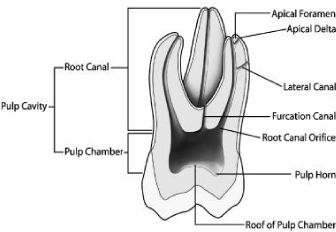
Figure 1: Vertical section of a permanent molar.
The number of blood and lymphatic vessels, and associated nerves decrease gradually. Endodontic treatments are rather easy to operatein young dental pulp, whereas such therapies are becoming more difficult in the elderly, especially in the root part of the tooth. This implies that the apical closure and volume restriction have to be gradually taken into account. At the root furcation, the pulp chamber becomes thicker, with a series of successive laminations. The lateral walls enlarge and become broader and thicker. At the roof level, reactionary dentin is strongly influenced by pulp aging. The overall restriction of space also contributes to the process of aging [4].
The young pulp chamber includes mainly (1) the floor (or furcation zone, located between the roots), (2) the occlusal wall or roof, and (3) reactionary dentin formed along the sidewalls (mesial or distal aspects) or lateral walls. Again, the volume of the aging pulp is reduced on the occlusal direction (reduction in height), and more than in the mesiodistal diameter. Reparative dentin is formed in case of a horn exposure. Reactionary dentin is more physiological and forms the fastest on the floor of the pulp chamber. Secondary dentin is formed at a slower rate on the occlusal wall (or roof), and far less is formed (3) on the side or lateral walls [5] (Figure 2).
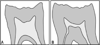
    Figure 2:  Difference in the pulp volume between a (A) young (7 years old)
and (B) old (55 years old) mandibular molar.
For these reasons, pulp regeneration occurs after rapid or slow carious lesion, after partial or total degradation (mechanical or infectious) of the pulp tissue. The objectives of this review on the root canal treatment diverges totally 1) according to the age of the pulp, 2) may be restricted to a simple pulp exposure or 3) follow the partial or total destruction of the pulp tissue. Reparative dentin may well be limited to the horn of the pulp, forming a dentinal bridge, with or without the formation of tunnels. Infectious bacteria may totally invade and degrade the dental pulp, leaving a residual tissue, a situation leading as end-point to the degradation of the root canal tissue. In the first case, pulp may be repaired, whereas in the second case, regeneration allows the regeneration repair of the teeth.
Species variations are detectable and they have also to be taken into account when endodontic treatments are carried out. For example, in rat’s molar deposition of reactionary dentin is the reverse of what is seen in humans. The reactionary dentin formation occurs fast (1) in the lateral walls, whereas there is a decrease in the (2) occlusal roof, and even more (3) at the furcation area [6]. Pulp stones are classified into three different zones: true or false denticles, located near or just in the central part of the pulp, or appear as widely spread diffuse mineralization’s. With time, pulp stones add to endodontic difficulties.
In addition to pulp stones, when the dental pulp becomes older, reactionary dentin deposition is detected along the lateral walls, whereas the reverse is observed in the young pulp. Roof dentin formation is slow, and less reparative dentin is formed in the furcation zone. Again, the overall pulp volume is decreased, but not the mesiodistal width that remains stable. Pulp horns are permanent structures, despite the general decrease in pulp volume and the asymmetric closure of the pulp.
Changes in the composition of the dental pulp
The pulp cellular components decrease in number in the elderly. Pulp fibroblasts or pulpoblasts [7] constitute the bulk of pulp cells. They have a high content of Rough Endoplasmic Reticulum (RER), and prominent Golgi apparatus. Pulpoblasts are linked by intercellular junctions (desmosome-like and gap junctions), favoring cell sliding from the center toward the periphery of the pulp. Pulp cells are implicated in the synthesis and secretions of type I and III collagens, glycosaminoglycans, and other collagenous and noncollagenous extracellular matrix components.
The dental pulp is equipped with inflammatory cells, e.g. other cellular components: peripheral T cells (helper/inducer and cytotoxic/ suppressor), and dendritic cells located primarily in the odontoblastic layer. They present HLA-DR antigens on the cell surface to CD4+ T-lymphocytes. Other antigen-presenting cells are located in a more central portion of the pulp. Class II antigen activated macrophages are 4 times more common than the dendritic cells. In the normal (non-inflammatory) pulp, there are no B cells [8].
Effect of age on pulp tissue: Nerve and blood supply of the dental pulp becomes more fibrous and less cellular. Mediators of inflammation include lymphocytes, plasma cells and macrophages. Non-specific mediators such as histamine, bradykinin, serotonin, interleukins (IL-1 and IL-2) are also released within the pulp. Mast cells, the main source for histamine is found in the inflamed pulp. A fourfold increase in pulp histamine levels is found within 30 minutes of thermal injury, supporting that this mediator may play a role in pulp inflammation. Plasma or tissue kallikreins lead to the production of bradykynin producing signs of inflammation. Various prostaglandins, thromboxanes and leukotrienes result from inflammatory processes [9]. Platelets aggregated in the vessels. Platelets aggregated in the vessels are releasing serotonin and vascular recruitment, as well as inflammatory processes [10]. They release serotonin from platelets membranes and the resulting mediators consist in the release of various mediators involved in the recruitment and differentiation of stem cells and reparative processes.
Microcirculation: In the pulp, arterioles feeding individual microcirculation and shunt vessels are observed. Their organization included arterio-venous anastomoses, venous-venous anastomoses, or U-turn loops bypassing the capillary bed. Capillaries irrigate successive 30 μm areas near the outer limits of the coronal pulp chamber. In the root, a fisher-like network is seen. Vascular thrombus blocks any potential for the radicular pulp repair, leading to tissue degradation.
Sensory nerves: They are branches arising from the maxillary and mandibular divisions of the trigeminal nerve. Nerve plexus contains both large myelinated A-d and A-β fibers (2-5 μm in diameter) and smaller unmyelinated C fibers (0.3-1.2 μm). Nerves end beneath the odontoblast layer, forming the Raschkow plexus. Nerves are present in the inner dentin, in the 150 μm layer located at the dentin-pulp border, but not further in the more external outer dentin.
Evaluation of a successful root canal treatment
Combining the healing/prevention of disease (apical periodontitis) and understanding the risk factors associated with endodontic failure allows increasing the chance of success of a Root Canal Treatment (RCT). It depends of the accuracy of the initial diagnostic and planning, the excellence of disinfection, instrumentation and filing procedures (antimicrobial strategies, root canal shaping and coronal and apical seal), and finally of the rehabilitation management allowing determining if a root canal treatment is a success or a failure. Constant or intermittent pain, and discomfort may suggest an endodontic failure. Reevaluation over time, absence or regression of apical periodontitis, may cast doubt on the reality of a RCT success [11].
Preparation of the root canal system includes both enlargement and shaping closely associated with disinfection [12]. They are referred to chemo-mechanical or bio-mechanical preparation. The aims of this mechanical root canal preparation are seven. 1- The removal of vital and necrotic tissue from the main root canal(s), 2- The creation of sufficient space for irrigation and medication, 3-Preservation of the integrity and location of the apical canal anatomy, 4- Avoidance of iatrogenic damage to the canal system and root structure, 5-Facilitation of canal filling, 6-Avoidance of further irritation and/ or disinfection of the peri-radicular tissues, 7- Preservation of sound root dentine to allow long-term function of the tooth. The various techniques include manual preparation, automated root canal preparation, sonic and ultrasonic preparation, use of laser systems, and Nickel-Titane (NiTi) instruments. Apical extrusions of debris are held responsible for post-operative flare-ups and bacteremia.
RCT should contribute primarily to the bone healing of an apical lesion. Apical lesions appear either as an unlimited diffuse radiolucent area around or near the apex, or as granuloma or cyst included inside or limited by a pouch. Histological investigations have demonstrated either dentinoclastic resorptions, visible only after tooth extraction and examination with the Scanning Electron Microscope (SEM). Granuloma or apical lesions are very similar radiographically, and no difference can be established on a simple visual basis. Treatments of the root canal are conclusive when they lead to bone healing. Bone gradually is reformed, trabeculae reappear, radiographic density is increase and no evidence of infection is detectable. The root canal treatment is a success if it is leading to clear visualization of an apex. In many cases after RCT, the apex is forming and elongating, with a growth in length of the root. Cementum apparently is formed, which is indeed a cellular cementum. The periodontal ligament, well known to contain also stem cells, is also healed. The tooth becomes again functional (Figure 3).
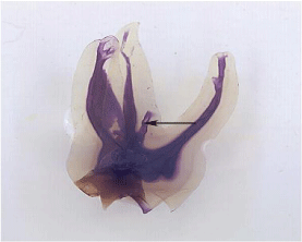
Figure 3: The dental pulp of a molar.
Old pulps are described as being “sclerosed” or “calcified”. Pulp space diminishes through life by the deposition of secondary dentin. This occurs commonly in pulp horn, and in the floor and roof of the pulp chamber of molars, converted from a large rectangular cavern in the young to a flat disc in the elderly (Figures 2-4). Pulp space is further reduced by reactionary and reparative dentine formation, formerly classified as tertiary or irritation dentine. Calcified inclusions are forming pulp stones, concentric spheroidal masses of calcified material. Between such masses, blood vessels and nerve cells are foci for linear calcified deposits. Prevention or healing of apical periodontitis supposes that microorganisms spread in large numbers and invade the periradicular tissues after local anesthesia followed by the canal aperture. Files can now be active with lubricants working with Ethyleneamine Tetracetic Acid (EDTA). Copious irrigation/ lubrication. “Apical calcification” is a rare event in a canal system, and despite efforts, canals are not negotiated to the definitive length. In conclusion, endodontics can be achieved in the elderly, in accordance with an actual diagnosis, good radiographs and overcoming the challenges posed by calcification of the root canal system [13].
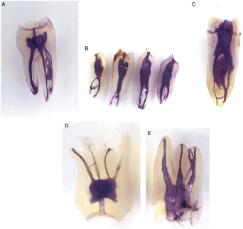
Figure 4: Premolar and molar dental pulp.
Ten properties of an ideal root filling material have been listed by Grossman [14]. An ideal root filing 1-Should be easily introduced into the canal, 2- It should seal the canal laterally as well as apically, 3- It should not shrink after being inserted, 4- It should be impervious to moisture, 5- It should be bacteriostatic or at least not encourage bacterial growth, 6- It should be radiopaque, 7- It should not stain tooth structure, 8- It should not irritate periapical tissue, 9- It should be sterile, or easily sterilized before insertion, 10- It should be easily removed from the root canal if necessary. Gutta-percha, silver points, resin-based core filling materials were used as core materials. As sealers, chloro-percha dissolved in chloroform, zinc-oxide-eugenol based sealers, glass-ionomer-based sealers, materials with calcium hydroxide, pass successfully antibacterial testing. They did not show any neurotoxicity, or leakage in the crown or in roots. Root canal treatments are a success when the contours, width and structure of the periodontal ligament are normal, even around the excess filling. It is a failure of endodontic treatment when there is a decrease of the peri-radicular rarefaction, or an unchanged bone density rarefaction, or an increase from the initial lesion [15].
To summarize, mechanical preparation may result in a significant reduction of bacteria, but will not leave bacteria-free root canals. Mechanical preparation should be assisted or completed by appropriate irrigants and intra-canal medicaments [12].
Endodontic treatment for children
Dental trauma leads often to pulpal necrosis (with a prevalence being about 4-59% of the incisors). The short roots increase the risk of subsequent fracture [16]. Healing of apical periodontitis, promotion of continued root development and restoration of the functional pulpal tissue are crucial for the regeneration of a functional pulpal tissue.
MTA apexification seems to offer superior advantages over the traditional Ca(OH)2 method. The characteristics of Ca(OH)2 as an intracanal medicament over in vitro studies related to the antimicrobial effects are as the following: hydroxyl ions are released, and antimicrobial effects against endodontic pathogens are efficient [17]. However, human clinical studies showed limited antimicrobial effects, microorganisms being reduced but not eliminated. Some species has resistance to Ca(OH)2. This is actually the case for Enterococcus faecalis, and/or Candida albicans. One-visit treatment is apparently as effective as a two-visit treatment, with interappointment intracanal medicaments.
Stem cells are found in the dental pulp, in the apical papilla and in inflamed periapical tissue. A massive influx of mesenchymal stem cells is found into the root canal space. These cells may contribute to regenerate a dental pulp. Growth factors may also contribute to guide the differentiation of cells into the pulp-dentin complex. The scaffold seems also to be important, either as endogenous scaffold (collagen, dentin) or as synthetic material (hydrogel, MTA and other compounds). Differences may occur between regeneration and revascularisation. These differences may play a role in pulp repair and in the selection of appropriate treatments to patients.
Isolation with rubber dam is a crucial step. Pediatric endodontic treatment should be directed toward pulpotomy rather than pulpectomy [18]. Enamel and dentine are thinner in temporary tooth than in permanent teeth. The pulp chambers with extended pulp horns are larger in the deciduous teeth. Either the root canal is treated or deciduous teeth should be extracted. It is quite clear that extraction should be avoided in case of bleeding disorders or medical conditions where anesthesia is contra-indicated. In other cases, endodontic treatments of primary teeth are indicated, especially in the presence of pulp necrosis or pulpitis.
Indirect pulp capping may provide some results after lining the cavity with a thin layer of calcium hydroxide, stimulating the formation of secondary dentine. Antibiotic or anti-inflammatory drugs may also be used, however pulp necrosis and abscess formation result without significant symptom during the initial steps. A dentinbonding agent, resin modified glass-ionomer, calcium hydroxide; zinc oxide/eugenol may be placed for indirect capping over the remaining carious dentin to stimulate healing and repair. The seal prevent microleakage. Interim Therapeutic Restorations (ITR) may be used for caries control in teeth with carious lesions that exhibit sign of reversible pulpitis. Reparative dentin protects the pulp. Indirect pulp capping has higher success than pulpotomy in long-term studies. Therefore, indirect pulp treatment is preferable to a pulpotomy when the pulp is normal and displays a diagnostic of reversible pulpitis.
Direct pulp capping is recommended when a small traumatic exposure occurs during cavity preparation in a vital non-infected pulp. Placed directly over the pulp, as a liner, undifferentiated mesenchyme-like cells differentiate into osteoclastic cells leading to internal resorption. Vital pulpotomy techniques may also be used. Under local anesthesia, the coronal pulp chamber is opened, removed by using a large excavator or a sterile bur cooled with sterile water or saline. Hemostasis is obtained by compression with a sterile cotton wool. Tables 1-3 summarize the major methods used during endodontic treatment of deciduous pulps. Buckley’formocresol is placed over the radicular pulp for 5 minutes, to fix the inflamed tissue and bacteria. It allows healing of the unaffected pulp. When the hemorrhage is completely stopped, zinc oxide- eugenol or glass ionomer cement is applied, and the tooth restored. Paraformaldehyde and Beechwood creosote are also useful tools.
    Tricresol
    35%


    Formaldehyde
    19%


    Glycerol
    15%


    Water
    31%
Table 1: Buckley’s formocresol.
    Parformaldehyde
    1 g


    Carbowax 1500
    1.30 g


    Lignocaine
    0.06 g


    Propylene glycol
    0.6 ml


    Carmine
    10 mg
Table 2: Paraformaldehyde devitalizing paste.
    0-Methoxy phenol (Guaicol)
    47%


    P-Methoxy phenol
    26%


    2-Methoxy, 4 methyl phenol    (Cresol)
    13%


    M-Methoxy phenol
    7%


    Other
    7%
Table 3: Beechwood creosote.
Pulpotomy
Pulpotomy may be performed in a primary tooth with extensive caries, without evidence of radicular pathology. The coronal pulp is amputated, and the remaining radicular pulp is treated with a longterm clinically successful medicament such as Buckley’s solution of formocresol or ferric sulfite. The objective is then to remove the carious dentin, leaving the carious mass over the pulp. The decrease of bacteria and the aim is to arrest the caries development. The placement of a final well-sealed restoration involves either in one appointment caries excavation and the formation of tertiary dentin; or a step-wise technique is used based on patient circumstances. If there is no symptom of pulpitis the pulp is judged to be clinical and radiographically vital and able to heal from the carious insult.
Translational opportunities to use strategies from basic stem cells research into practice imply a cascade of methods in endodontics. Step by step it presumably involves indication/clinical diagnostic, disinfection/irrigation, and root canal preparation/pretreatment. They generate new insight into pulp regeneration mechanisms [19].
If the necrosis is limited to the crown, the one-stage technique has to be used. If the root canal has to be cleaned, shaped and irrigated, the two-stage technique is required (Figures 5,6).
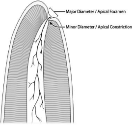
Figure 5: Apical part of a root: apical foramen and constriction.
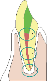
    Figure 6:  In 1: coronal leakage; in 2 Place where surviving microorganisms
are found, and 3 Apical zone preventing accumulation of stagnant fluids.
The immature root with a necrotic pulp and apical periodontitis implies that the infected root canal space cannot be disinfected with the standard root canal protocol, endodontic files being aggressive for the periapex. Namely, there is no barrier for stopping the root canal material. In addition, the roots of these teeth display higher susceptibility to fracture [20].
These teeth are infected, and the first phase of the treatment is to disinfect the root canal system. After access to the canal is made, a file is placed to this length, and confirmed radiographically. Copious irrigation with 0.5% sodium hypochlorite or less are used. The canal is dried and filled with a tri-antibiotic paste (metronidazole, ciprofloxacin and minocycline). Cleaning, shaping and intra-canal medication (calcium hydroxide or mineral trioxide aggregate) are performed at that stage in a standardized manner [21]. Calcium hydroxide is packed against the apical soft tissue with a plugger to initiate hard tissue formation. At 3 months a radiograph is taken to evaluate the hard tissue barrier formed, probed with an instrument. It takes between 6 and 18 months (4 to 13 months according to some authors) to evaluate if the barrier is complete enough to provide a stop to a filling material (Figures 7,8).
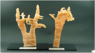
    Figure 7:  Apical part of a molar root canal (to the left) and canine (to the
right).
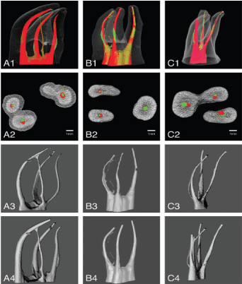
    Figure 8:  3D reconstitution (MicroCT or cone beam) of the canal systems a
maxillary molar.
Mineral Trioxide Aggregate (MTA) barrier may be used. A collagen-enhanced scaffold stimulates the revascularization process [22]. Discussion between revascularization and pulp regeneration is still open, focusing on the formation of a periodontal ligament, and stimulating pulp regeneration from the pluripotential cells in the periapical region. Apexogenesis is also a crucial point and pulp preservation is a primary goal for the young permanent dentition. Revascularization is a treatment for permanent teeth with necrotic pulp and incomplete root development [23]. Treatment of immature permanent teeth with necrotic pulp, with or without apical pathosis, implies the risk of dentin wall fracture, extending gutta percha into the periapical tissue during the compaction of root canal filling. Disinfection of the root canal and stimulation of stem cells can induce the formation of new hard tissue and continue the root development. Irrigants and medicaments have been chosen for their antimicrobial effects without consideration for their effects on stem cells and to the dentinal microenvironment [24].
Permanent dentition
Immature permanent tooth: After a trauma or an infection, pulp may be non-vital with or without an apical periodontitis. The successive endodontic steps imply the opening (access) of the pulp chamber with suppression of overhanging tissues (enamel and dentine) and manage the access opening. A rubber dam isolates the tooth from the bacterial environment and saliva contamination. Then, the next step needs root canal access, cleaning, disinfecting, and shaping (enlarging). Irrigating solution such as NaOCl disinfect the content of the root canal. In some cases, hypochlorite accident is a well-known complication that may occur during canal therapy.
Endodontics treatment maintains the health and well being of the elderly. The pulpo-dentinal complex undergoes calcific change in the pulp.
Pulpectomy in conventional root canal treatment for exposed, infected, and/or necrotic teeth aims to eliminate pulpal and periradicular infection. The roof of the pulp chamber is eliminated. Debridement, disinfection and shaping of the root canals after elimination of the crown and root canal system is accomplished with a biologically acceptable, non-resorbable filling material (gutta percha or other materials)
The aim of such therapies is to fill the main canal and secondary canals. The procedure should include root end closure (apexification) and the formation of an apical barrier is confirmed by clinical and radiographic evaluation [25]. Bioactive nanofibrous scaffolds, injectable scaffolds and stem cells lead to stem cell-based strategies [26]. Immature necrotic permanent teeth treated by mechanical instrumentation do not prevent further weakening of already thin and fragile root dentinal walls. Root canal irrigations associated with antibiotic pastes composed of metronidazole, ciproxacin, and minocycline or calcium hydroxide has been used. Antibiotic pastes at clinical concentrations affect the survivability of SCAPs.
Sodium hypochlorite (NaOCl), chlorhexidine gluconate (CHX), 17M EDTA, citric acid, MYAD and 37% phosphoric acid solution were used for a successful disinfection and cleanness of the root canals. CHX is an irrigative substance. It possesses a wide range of antimicrobial activity, lower toxicity than NaOCl whilst demonstrating efficient clinical performance, lubricating properties, rheological action. It inhibits metalloproteinases, is chemically stable, odorless, and water-soluble [27]. In case of open apex, root resorption, foramen enlargement and root perforation or in case of allergy due to bleaching solutions CHX should be avoided. It may be used as intra-canal medicament and diffusion into the dentinal tubules. CHX gel reduces the smear layer formation, a phenomenon that does not occur with the liquid form. CHX effect at low concentration is a bacteriostatic effect. At higher concentration, CHX has a bactericidal effect due to the precipitation and/or coagulation of bacterial cells, caused by protein cross-linking, resulting in cell death, and leaving cell debris in the root canal. In a few cases, possible adverse effects have been reported, including cytotoxicity and genotoxicity [28]. In vitro cytotoxic effects are dose dependent on human osteoblastic cells. Mutagenicity in microorganisms does not eliminate bacterial spores, and cytotoxicity (related to the potential damage of certain substances to the DNA) has been reported. Adverse effects have been related to topical or oral applications, including reversible discolorations, transient taste disturbances and burning sensation of the tongue. From the published data, it may be concluded that the biocompatibility of CHX is acceptable.
Prospects in endodontic treatments
After root decontamination with antimicrobial irrigants solutions followed by the insertion of a bioactive scaffold containing growth factors and/or stem cells, a new pulp tissue containing odontoblasts had the potential of inducing the formation of dentin-like tissue. This new pulp tissue increases the thickness of dentin walls and provides re-establishment of tooth functions in the oral cavity.
The success of single versus multiple visits for endodontic treatment of permanent teeth (Root canal Treatment - RCT) is characterized by the absence of symptoms and clinical signs in a single visit or over two or more visits, with or without medication. No difference was found between a single visit and multiple-visit treatments in terms of radiological failure, immediate post-operative pain, swelling or flare-up incidence, and fistula formation. There is no evidence to suggest that one treatment is better than the other. The single visit treatment implies a higher frequency of swelling, and late postoperative pain [27]. Similar healing might be obtained through one- and two-visit antimicrobial treatment. Most short- and long-term complications are similar in terms of frequency. Root canal irrigants such as sodium hypochlorite and chlorhexidine are effective at reducing bacterial cultures [29].
Pulp-dentin regeneration
Induced pulpal mesenchymal stem cells accelerate the trans differentiation capacities of pulp cells into functional odontoblasts when transplanted into the root canal microenvironment. Dental pulp stem cells of permanent and temporary teeth, apical papilla stem cells, periodontal ligament stem cells and cells obtained from the dental follicle may provide tools implicated in pulp healing and regeneration. DPSCs were isolated and characterized from dental pulp tissue. They may also be obtained from the apical papilla, exfoliated deciduous teeth, and from the Periodontal Ligament (PDL). Along this line of evidences, Bone Mesenchymal Stem Cells (BMMSCs) multiply and display limited expansion. They express the Oct-4, Nanog, STRO- 1, CD 73, CD90, CD105 and CD146. They are negative for CD14, CD34, CD45 and human leukocyte antigen-DR. The combination of BMSCs with oral epithelial cells from rat embryo generates toothlike structures, expressing Dentin Sialophosphoprotein (DSPP) and Dentin Matrix Protein 1 (DMP1). Inductive materials promote the differentiation of stem cells into SCAPs. Pulp regeneration may be obtained by capping the horn of the dental pulp with calcium hydroxide, mineral trioxide aggregate and dentin-like mineral structures. Revascularization and angiogenesis, neurogenesis, and apexogenesis are parts of the future of valid endodontic treatments [30,31].
Conclusion
Substantial differences between young and old dental pulp lead either to pulp repair including the formation of reparative dentin, filling of the pulp exposure with a calcified dentinal bridge, followed by the apexification of the tooth, or to pulp regeneration by activation and proliferation of residual stem cells. Pulp regeneration is pursued by the mineralization of the pulp tissue, which appears to be in the next future the most favorable endodontic treatment.
References
- Kuboki Y, Takagi T, Sasaki S; Saito S, Mechanic GL. Comparative collagen biochemistry of bovine periodontium, gingiva, and dental pulp. J Dent Res. 1981; 60: 159-163.
- Nielsen CJ. Collagen changes in dental pulp Front oral Physiol. 1987; 6: 111-125.
- Shuttleworth CA, Berry L, Kielty CM. Microfibrillar components in dental pulp: Presence of both type VI collagen- and fibrillin-containing microfibrils. Archs Oral Biol. 1992; 37: 1079-1081.
- Vertucci FJ. Endodontic Topics. 2005; 10: 3-29.
- Sicher H. Mosby Company Saint Louis. 1966 Chapter V Pulp. 127-154.
- Salomon JP, Lecolle S, Roche M, Septier D, Goldberg M. A radioautographic comparison of in vivo 3H proline and 3H serine incorporation in the pulpal dentine of rat molars: variations according to the different zones. J Biol Buccale. 1990; 18: 307-312.
- Baume LJ. The biology of pulp and dentine. A historic, terminologic-taxonomic, histologic- biochemical, embryonic and clinical survey. Monogr Oral Sci. 1980; 8: 1-220.
- Goldberg M, Njeh A, Uzunoglu E. Is pulp inflammation a prerequisite for pulp healing and regeneration. Mediators Inflamm. 2015; 2015: 347649.
- Yu C, Abbott PV. An overview of the dental pulp: its functions and responses to injury. Aust Dent J. 2007; 52: S4-S16.
- Baudry A, Alleaume-Butaux A, Dimitrova-Nakov S, Goldberg M, Schneider B, Launay JM, et al. Essential roles of dopamine and serotonin in tooth repair: functional interplay between odontogenic stem cells and platelets. Stem Cells. 2015; 33: 2586-2595.
- Estrela C, Holland R, Estrela CRA, Alencar HG, Sousa-Neto MD, Pécora JD. Characterization of successful root canal treatment. Brazilian Dent J. 2014; 25: 3-11.
- Hülsmann M., Peters OA, Dummer PMH. Mechanical preparation of root canals: shaping goals, techniques and means. Endodontic Topics. 2005; 10: 30-76.
- Finnbarr P, Whitworth JM. Endodontic considerations in the elderly. Gerodontology. 2004; 21: 185-194.
- Grossman LI. Endodontic Practice. Philadelphia: Lea & Febinger, 1978.
- orstavik D. Materials used for root canal obturation: technical, biological and clinical testing. Endodontic Topics. 2005; 12: 25-38.
- Hargreaves KM, Diogenes A, Teixeira FB. Treatment options: biological basis of regenerative endodontic procedures. J Endod. 2013; 39: S30-S43.
- Kim D, Kim E. Antimicrobial effect of calcium hydroxide as an intracanal medicament in root canal treatment: a literature review- Part II. In vivo studies. Restor Dent Endod. 2015; 40: 97-103.
- Carrotte P. Endodontic treatment for children. Br Dent J. 2005; 198: 9-15.
- Peter OA. Translational opportunities in stem-cell-based endodontic therapy: where are we and what are we missing? J Endod. 2014; 40: S82-85.
- Trope M. Treatment of the immature tooth with a non-vital pulp and apical periodontitis. Dent Clin N Am 2010; 54: 313-324.
- Maniglia-Ferreira C, de Almeida Gomes F, Guimaràes NLS, Vitoriano MM, Ximenes TA, de Souza BC, et al. Endodontic treatment for necrotic immature permanent teeth using MTA and calcium hydroxide. A retrospective study. RSBO. 2013; 10: 116-121.
- Banchs F, Trope M. Revascularization of immature permanent teeth with apical periodontitis: new treatment protocol? J Endod. 2004; 30: 196-200.
- Wigler R, Kaufman AY, Steinbock N, Hazan-Molina H, Torneck CD. Revascularization is a treatment for permanent teeth with necrotic pulp and incomplete root development. J Endod. 2013; 39: 319-326.
- Diogenes AR, Ruparel NB, Teixeira FB, Hargreaves KM. Translational science in disinfection for regenerative endodontics. J Endod. 2014; 40: S52-S57.
- Clinical Affairs Committee, American Academy of Pediatric Dentistry. Guideline on Pulp Therapy for Primary and Immature Permanent Teeth Clinical Practice Guidelines. 244- 252.
- Albuquerque MTP, Valera MC, Nakashima M, Nör JE, Bottino MC. Tissue-engineering-based strategies for regenerative endodontics. J Dent Res. 2014; 93: 1222-1231.
- Manfredi M. Figini L, Gagliani M, Lodi G. Single versus multiple visits for endodontic treatment of permanent teeth. Cochrane Database Systamatic Review. J Endod. 2008; 34: 1041-1047.
- Gomes BPFA, Vianna ME, Zaia AA, Almeida JFA, Souza-Filho FJ, Ferraz CCR. Chlorhexidine in endodontics. Brazilian Dent J. 2013; 24: 89-102.
- Fedorowicz Z, Nasser M, Sequeira-Byron P, de Souza RF, Carter B, Heft M. Irrigants for non-surgical root canal treatment in mature permanent teeth. Cochrane Database Sys Rev 2012; (9): CD008948.
- Cao Y, Song M, Kim E, Shon W, Chugal N, Bogen G, et al. Pulp-dentin regeneration: current state and future prospects. J Dent Res. 2015; 94: 1544-1551.
- Li Y, Shu L-H, Yan M, Dai W-Y, Li J-J, Zhang G-D, et al. Adult stem cell-based apexogenesis. World J Methodol. 2014; 4: 99-108.