
Research Article
J Dent & Oral Disord. 2018; 4(1): 1084.
A New Type of Radiographic Template for Presurgical Radiographic Examination in Implant Restorations
Kourtis S*
Department of Prosthodontics, National and Kapodistrian University of Athens, Greece
*Corresponding author: Stefanos Kourtis, Associate Professor, Department of Prosthodontics, National and Kapodistrian University of Athens, Greece
Received: December 18, 2017; Accepted: January 31, 2018; Published: February 08, 2018
Abstract
Various radiopaque materials have been used as radiopaque markers for radiographic templates. A new type of diagnostic template is presented in this article that allows radiographic imaging of the intended implant axis and the contour of the restoration in CT Dental Scan and Cone Beam Computer Tomography (CBCT). The use of amalgam powder as a coverage layer on the outer surface of a template offers detailed imaging of the restoration contour in correlation to the bone substrate. The diagnostic template can be transformed easily to an accurate surgical template.
Keywords: CT- Dental scan; CBCT; Radiographic guide/ template, Restoration contour; Dental implants
Introduction
Implant restorations need detailed presurgical planning and radiographic control. CT-Dental Scan and Cone Beam Computer Tomography (CBCT) are used extensively nowadays in the clinical practice, in order to investigate the bone substrate before implant surgery [1-4]. The most common types of diagnostic templates are constructed from autopolymerizing resin with radio-opaque materials used as markers to determine specific anatomical areas. As radio-opaque markers various materials have been used, including metal rods, titanium rods, guttapercha, barium sulfate etc [4-10].
The aim of this paper was to describe a new type of diagnostic template for CT- Dental Scan or CBCT that allows imaging of the restoration contour in relation to the bone substrate.
Technique
Study casts are mounted on a semi-adjustable articulator and a wax-up is made on the area to be restored. In cases of edentulous patients the diagnostic set-up or the existing denture (if satisfactory, as in the presented case) can be used. From the wax-up (or the existing denture) silicone impressions are made and a duplicate from heat polymerizing clear acrylic resin is fabricated. Alternatively autopolymerizing resin can be used (Figure 1).
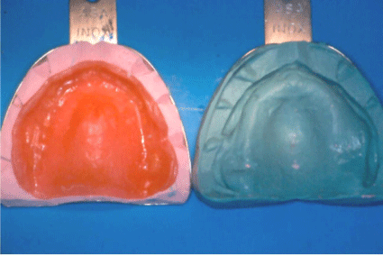
    Figure 1:  The existing denture is duplicated using silicone impression
material.
The acrylic teeth in the duplicate denture are drilled in the center (3 mm diameter) in a parallelographing milling device, along the desired axis of the implant. If the drillings are not necessarily parallel, as in the edentulous maxilla, a line is drawn along the axis of the tooth in the labial area with a marker and the drilling can be accomplished manually parallel to the markings. The parallel axis of the drillings can be checked visually by inserting laboratory rotating instruments in the drillings (Figure 2). The hollow space created by the drilling is filled with guttapercha (Figure 3). Alternatively titanium rods can be used as they do not cause artifacts on the CT image.

    Figure 2:  Lines are drawn along the teeth axes and the drillings are performed
parallel to the markings.
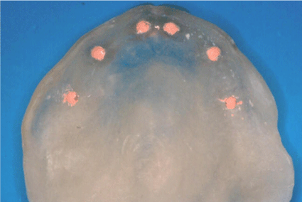
Figure 3: The hollow spaces of the drillings are filled with guttapercha.
The outer contour of the acrylic teeth is covered with amalgam powder dissolved in transparent nail varnish. The varnish is applied with a soft brush as a thin layer on the whole surface (labially and palatally) up to the cervical areas (Figure 4). The varnish layer should be restricted to the areas where implants are planned to avoid unnecessary image “noise” and to facilitate area recognition on the cross-section images of the Computer Tomography.
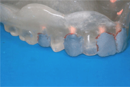
    Figure 4:  The outer surfaces of the template on specific teeth are covered
with amalgam powder dissolved in transparent nail varnish.
The patient is then referred to perform a radiographic examination, CT Dental Scan or CBCT. On the examination of the reconstructed cross-section images the radiopaque markers can be easily identified in the specific areas and also the contour of the acrylic teeth. In this way the desired implant axis can be correlated to the existing bone substrate and the contour of the planned restoration (Figure 5,6).
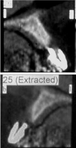
    Figure 5:  Cross-section images of the anterior maxillary teeth. The
guttapercha marker can be distinguished within the radiopaque contour from
the amalgam powder.

    Figure 6:  CBCT cross-section image at the premolar region of another
patient with the same type of markers.
The diagnostic template can be easily transformed to a surgical guide by removing the nail varnish with acetone and the guttapercha from the hollow spaces. In cases of edentulous patients (as in the presented case) the anterior palatal part of the template is removed to allow better surgical approach, maintaining the guiding labial surfaces (Figure 7,8).
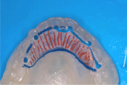
    Figure 7:  The anterior palatal part of the template will be removed for the use
as a surgical guide.
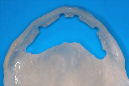
Figure 8: The template transformed to a surgical guide.
The use of a template reproducing the desired contour of the restoration at CT-Scan images may contribute significantly to the treatment planning and provide the clinician with valuable information. In cases with severe bone resorption in the anterior maxillary region, the correlation of the restoration contour to the existing bone substrate may lead the clinician to proceed to bone augmentation or to offer to the patient a removable restoration (Figure 9,10).
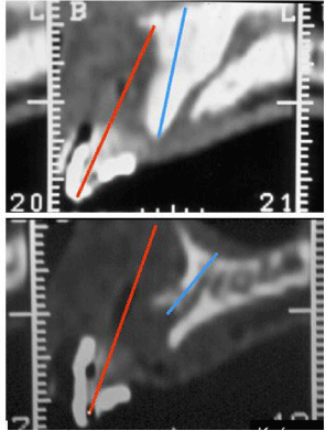
    Figure 9:  Cross-section images of the anterior maxillary region of another
patient with severe bone resorption. Amalgam powder has been used to
indicate the contour of the restoration and no guttapercha has been inserted.
Red line (left) indicates the prosthetically favorable implant axis and blue line
(right) the axis of the implant as imposed by the existing bone substrate. In
this case an augmentation procedure is necessary.
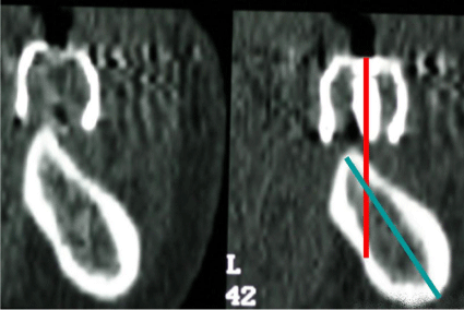
    Figure 10:  CBCT cross-section image of the posterior mandibular region of
a patient with the same markers as in Figs 10 and 11. The red line indicates
the prosthetically favorable implant axis and blue line the axis of the implant
as imposed by the existing bone substrate.
Discussion
Computer-Aided Examinations (CBCT or CT Dental Scan) offer great help in presurgical planning. Detailed imaging is necessary in order to determine the desired implant axis related to the bone substrate. The representation of the restoration contour offers also valuable information as the implant axis can be determined (or modified) according to the contour of the planned restorations [11,12]. The insertion of implants in prosthetically favorable position may prevent later problems and complications during the clinical function of the implants [13-15].
Various materials have been proposed as radiopaque markers in the drill holes as guttapercha, titanium rods and silicone. Other materials have also been used as radiopaque coverage for the outer surface of the template as zinc foil or guttapercha diluted in chloroform [16-18]. Barium sulfate has also been proposed as radiopaque material added in the mass of acrylic resin [19]. The image provided by the amalgam powder has been the sharper shown in the CT-Scan [20].
The presented technique is simple, cost- and-time effective and requires no additional equipment. It can also be applied chairside without any laboratory stages. The transformation to surgical template requires minimum additional time and offers increased accuracy for implant placement [21,22]. The possibility of recognizing the occlusal level of the patient has been shown to be important for the proper reconstruction of the CT-Scan and the resulting vertical cross-section images [23]. For this reason, it is important to use a radiographic template with radiopaque markers, especially in completely edentulous patients.
The digital technology has made tremendous progress allowing not only a precise pre-surgical evaluation of the bone substrate but also offering the possibility of a completely digital planning and guided surgery [24,25]. On the other side it is not always affordable for the patient to undertake the cost of guided implant placement and an accurate presurgical examination using CBCT with a radiographic template may offer valuable information for the further treatment.
References
- Lekholm U. Clinical procedures for treatment with osseointegrated dental implants. J Prosth Dent. 1983; 50: 116-120.
- Parks ET. Computer ?omography applications for Dentistry. Dental Clinics of North America. 2000; 44: 371-374.
- Weinberg LA. CT Scan as a radiological data base for optimum implant placement. J Prosth Dent. 1993; 69: 381-385.
- Smith JP, Borrow JW. Reformatted CT for implant planning. Oral Maxillofac. Surg. Clin. North Am. 1991; 3: 805-825.
- Borrow JW, Smith JP. Stent markers materials for computerized tomograph assisted implant planning. Int J Periodont Rest Dent. 1996; 16: 60-67.
- Kraut R. Radiologic planning for dental implants; in: Block U S., Kent J N., Guerra, L R.: “Implants in Dentistry”. Chapter 5. W. B. Saunders Co., Philadelphia 1997; 33-44.
- Abrahams JJ. The role of diagnostic imaging in dental Implantology. Radiol Clin North Am. 1993; 81: 163-180.
- Ganz S. CT-Scan technology: An evolving tool for avoiding complications and achieving predictable implant placement and restoration. Int Mag Oral Implantol. 2001; 1: 6-13.
- Rosenfeld AL, Mecall RA. Using Computerized Tomography to develop realistic treatment objectives for the implant team; in: Nevins M., Melloning J T. (Edit): Implant therapy Vol. 2, Chapter 3. Chicago: Quintessence Publ Co. 1998; 29-46.
- Rothman SLG. Dental applications of Computerized Tomography. Quintessence Publ Co Chicago. 1998: 31-35.
- Mecall RA, Rosenfeld AL. influence of residual ridge resorption patters on implant fixture placement and tooth position. 1. Int J Periodont Rest Dent. 1991; 11: 8-23.
- Mecall RA, Rosenfeld AL. The influence of residual ridge resorption patters on fixture placement and tooth position in the partially edentulous patients. 2. Presurgical determination of prosthesis type and design. Int J Periodont Rest Dent. 1992; 12: 32-51.
- Lazzara RJ. Effect of implant position on implant restoration design. J Esthet Dent. 1993; 5: 265-269.
- Murel GA, Davis WH. Presurgical Prosthodontics. J Prosth Dent. 1988; 59: 447-452.
- Cehreli MC, Sahin S. Fabrication of a dual-purpose surgical template for correct labiopalatal positioning of dental implants. Int J Oral Maxillofac Implants. 2000; 15: 278-282.
- Tsuchida F, Hosoi T, Imanaka M, Kobayashi K. A technique for making a diagnostic and surgical template. J Prosthet Dent. 2004; 91: 395-397.
- Guerra LR, Finger IM. Principles of Implant Prosthodontics; in: Block, SK Kent, JN Guerra, L: Implants in Dentistry. Philadelphia: W. B. Saunders Co. 1997; Chapter 8, 74-85.
- Israelson H, Plemons JM, Watkins P, Sony C. Barium coated surgical stents and computer assisted tomography in the pre-op assessment of dental implant patients. Int J Periodont Rest Dent. 1992; 12: 52-61.
- Shahrabasi AH, Hansen CA. Surgical oral radiographic guide with a removable component for implant placement. J Prosthet Dent. 2002; 87: 330- 332.
- Kourtis S. Die fallbezogene praeoperative Bewertung und Behandlungsplannung. Teamwork. 2002; 15: 156-166 (in German, English abstract).
- Boskovic MM, Castelnuovo JC, Brudric JS. Surgical template for completely edentulous patients. Int J Period Restor Dent. 2000; 20: 181-189.
- Becker CM, Kaiser DA. Surgical guide for dental implant placement. J Prosth Dent. 2000; 83: 248-251.
- Kourtis S, Skondra E, Roussou I, Skondras EV. Presurgical planning in implant restorations: correct interpretation of Cone-Beam Computed Tomography for improved imaging. J Esthet Restor Dent. 2012; 24: 321-332.
- Angelopoulos C, Aghalloo T. Cone Beam Computed Tomography for the implant patient. Dent Clinic North Amer. 2011; 55: 141-158.
- Tsirogianis P, Pieger S, Pelekanos S, Kourtis S. Surgical and prosthetic rehabilitation through a complete digital workflow- A case report. Int J Comput Dent. 2016; 19: 341-349.