
Perspective
Austin J Dent. 2014;1(2): 1011.
Modifying Adult IOPA Film to Use in Children
Dr. Santosh Patil*
Assistant Professor, Department of Radiology, College of Dentistry, Al Jouf University, Sakaka, Saudi Arabia
*Corresponding author: Santosh Patil, Assistant Professor, Department of Radiology, College of Dentistry, Al Jouf University, Sakaka, Saudi Arabia
Received: October 15, 2014; Accepted: December 01, 2014; Published: December 03, 2014
Abstract
Dental radiographs are valuable diagnostic tools and expose the patient to minimal amounts of radiation. Periapical radiography describes intraoral techniques designed to show individual teeth and the tissues around the apices. Each film usually shows two to four teeth and provides detailed information about the teeth and the surrounding alveolar bone. In this article we describe a technique of converting adult film for using in children with an advantage of minimum costing.
Keywords: Intraoral radiography; Image receptors; Caries; Periapical
Introduction
X-rays were discovered in 1895 by Professor Wilhelm Conrad Roentgen. Dr. Otto Walkhoff is credited with the first dental radiograph [1]. The intraoral radiograph, when correlated with the case history and clinical examination, is one of the most important diagnostic aids available to the dental practitioner. Dental radiographs should be prescribed according to selection criteria guidelines and taken only for diagnostic and treatment purposes. Selection criteria guidelines are based on evidence of disease patterns and take into consideration the patient's medical and dental history, clinical signs and symptoms of disease, risk factors, age and dentition, and new or recall patient status [2]. When examined under proper conditions, diagnostic-quality intraoral radiographs reveal evidence of disease that cannot otherwise be found. They also play a major role in forensic identification. Intraoral dental radiographs fall into two main categories: bite-wing and periapical radiographs. The periapical view is taken of both anterior and posterior teeth. The objective of this type of view is to capture the tip of the root on the film. This is often helpful in determining the cause of pain in a specific tooth, because it allows a dentist to visualize the tooth as well as the surrounding bone in their entirety [3]. X-ray film of size 1 (24x40 mm) and 2 (31x41 mm) are used for taking radiographs of adults and 0 sized (22x35 mm) films are used for children.
Technique
This technique is useful for a child patient requiring multiple intraoral periapical radiographs for diagnosis and treatment. The routine size 2 IOPA film is taken and it is arbitrarily divided into two equal halves, then one half is covered with the 2 layers of lead foil which is present in IOPA film packet [Figure 1]. Then the film is placed in disposable plastic sleeves and positioned in patient's mouth (in the area where it is indicated) and exposed as per standard methods. Later the foil should be covered on the second half (exposed part) of the film and placed in the required quadrant and exposed. The film should be now processed using routine techniques and interpreted [Figure 2,3]. This technique may not be possible in children having shorter vestibular depth and limited mouth opening. There may be difficulty in indentifying the side of the radiograph by other dentists who have not exposed the film. The advantage of this technique is it is cost effective, it saves two third of price, as 0 size films are almost three time costlier than adult films. This technique is suitable for dental clinics with individual practitioners and not for hospitals and teaching institutes.
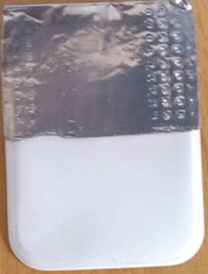
Figure 1: Adult IOPA film covered with lead foil.
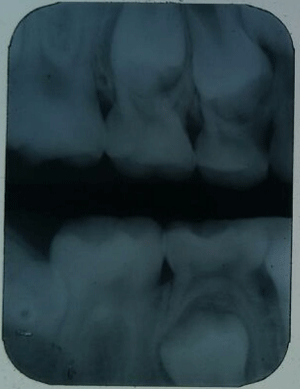
Figure 2: Image of posterior quadrants on single film.
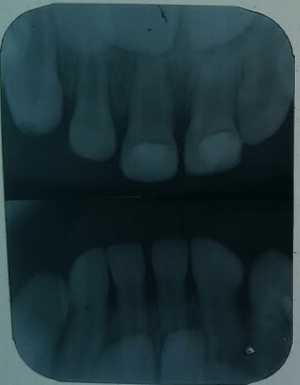
Figure 3: Image of anterior quadrants on single film.
References
- Pharoah MJ, White SC, eds. Oral Radiology: Principles and Interpretation. 4th edn. St. Louis: Mosby; 2001:49.
- American Dental Association and U.S. Department of Health and Human Services. The Selection of Patients for Dental Radiographic Examination, Revised 2004.
- Bird DL, Robinson D. Torres and Ehrlich modern dental assisting, 8th edn. St. Louis, Mo.: Elsevier, 2005.